
 New Zealand; International

News release
From:
Gisborne, New Zealand, April 8, 2025
The brain pulses with every heartbeat — how this impacts our understanding of the brain
New Zealand scientists at the forefront of global brain science breakthroughs with five papers involving Mātai and New Zealand teams published in UK Royal Society Journal.
The brain doesn’t just think — it moves, subtly pulsing with every heartbeat. Now, scientists from New Zealand and across the globe are exploring how these tiny movements and fluid shifts may hold the key to understanding brain health, ageing, and disease.
These ideas were the focus of the interdisciplinary Royal Society Pulsing Brain meeting, co-chaired by Associate Professor Samantha Holdsworth (Mātai and the University of Auckland), held in the UK.
The outcomes of this landmark meeting — organised by internationally distinguished researchers — have just been published as a dedicated issue of Interface Focus, the Royal Society’s premier interdisciplinary journal. The issue includes several international contributions highlighting recent breakthroughs in brain imaging.
Non-invasive ways to visualise and understand the brain’s movement
The opening perspective paper, “The Pulsing Brain: State of the Art and an Interdisciplinary Perspective”, explores how interdisciplinary approaches—spanning MRI, ultrasound, and mathematical modelling—are being used to non-invasively examine brain tissue pulsations and fluid flow. These insights are essential for advancing our understanding of brain health.
One exciting area of progress is advanced MRI technology, such as amplified MRI (aMRI), a technology spearheaded by the Mātai Medical Research Institute, which can make the brain’s micro-movements visible. These cutting-edge techniques provide a non-invasive window into the brain and have the potential to improve how we detect brain injuries, monitor ageing, and study neurodegenerative diseases like Alzheimer’s.
Four other major collaborative papers from Mātai, the University of Auckland’s Faculty of Medical and Health Sciences, the Auckland Bioengineering Institute, GE HealthCare, Ghent University, and other leading institutions are summarised in the following:
Impact of exercise on brain motion
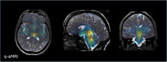
The brain moves with every heartbeat, and even mild exercise – like squeezing a handgrip – changes the way the brain moves. Quantitative-aMRI (q-aMRI), an advanced MRI technique developed in collaboration with Stanford University and Mātai, along with other standard MRI methods, were used to visualise brain dynamics during exercise.
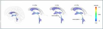
“During rest, the brain’s rhythmic motion matches the heartbeat, but it becomes less variable when the heart rate goes up during exercise. We were able to see that exercise reduces blood flow pulsatility (making the brain more stable) but increases cerebrospinal fluid movement. This means that faster heartbeats make brain motion more consistent” said researcher Jet Wright, a Mātai-based Auckland Bioengineering Institute PhD student.
“This could have implications for understanding concussion, brain pressure, and long-term neurological health. The study suggests regular movement or exercise may play a vital role in regulating brain fluid dynamics to keep the brain balanced and healthy. The research also opens up new possibilities for using exercise as a way to study brain health,” said senior author Dr. Eryn Kwon, Mātai Hugh & Moira Green Senior Research Fellow.
Brain blood flow changes as a marker for dementia and brain disease
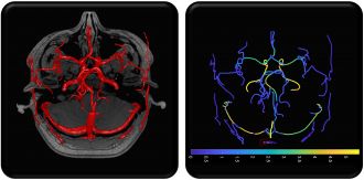
A study led by Sergio Dempsey from the Auckland Bioengineering Institute has found that a key measure of brain blood vessel stiffness – called pulse wave velocity (PWV) – does not increase with age in healthy people. PWV measures how fast blood pressure waves move through blood vessels, and faster waves usually mean stiffer vessels. 4D flow MRI is an advanced imaging technique that shows how blood flows through vessels in three dimensional space, over time (the fourth dimension). It gives a detailed picture of how fast and how smoothly blood travels through brain arteries. Using advanced 4D flow MRI, the research showed that in healthy individuals, brain PWV stays steady over time. This challenges long-held beliefs that brain blood vessels naturally stiffen as we age – something often linked to brain disease.
“These findings turn our understanding of brain ageing on its head, potentially highlighting the brains active protection to keep vessels more relaxed with ageing” said Sergio. The results raise important questions about how brain blood flow changes with age and what that means for brain health and diseases like stroke or dementia.
Understand fluid circulation in the brain with aMRI and computational modelling
One study, led by Sarah Vandenbulcke (Ghent University and Mātai visiting scholar), explored how aMRI could feed computational models to predict CSF movement more accurately, potentially improving diagnosis of fluid-flow disorders, such as hydrocephalous, increased brain pressure and other disorders.
New ways of analysing the brain’s movement
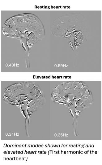
Research led by Haribalan Kumar (GE HealthCare) combined aMRI with Dynamic Mode Decomposition (DMD). DMD is a data analysis method that helps identify patterns and changes over time, enhancing the visualisation of brain motion synchronised to the heartbeat. This technique may enhance how we detect and monitor conditions like concussion or increased brain pressure.
“These studies and collaborations represent an advancement in non-invasive imaging and modelling enhancing our understanding of the brain’s mechanical environment. This progress opens new possibilities for diagnosis and treating brain conditions using motion-based biomarkers” said
Associate Professor Holdsworth.
About Mātai
Established in 2020, Mātai stands at the forefront of global medical research and innovation. Leveraging cutting-edge biomedical imaging technologies, artificial intelligence, and personalised medicine, we are dedicated to pioneering preventative and predictive healthcare solutions. With state-of-the-art facilities and collaboration with leading experts, our multidisciplinary team of world-class researchers is transforming scientific discovery. Our mission is to advance global healthcare using state-of-the-art MRI technology and personalised medicine for early and precise diagnostics and treatments.
Mātai contributes significantly to enhancing our understanding of health through MRI. MRI uses magnetic fields and radio waves to generate internal images of the body. MRI is safe, non-invasive, and can often reduce the need for unnecessary invasive surgeries. Advancing scan clarity, as well as shortening time needed for patients in the scanner, is transforming clinical research and diagnostic capability.
Multimedia
Attachments
Note: Not all attachments are visible to the general public. Research URLs will go live after the embargo ends.


