, size: c 1cm. Credit: Brigitte Schoenemann.")
Media release
From: Springer NaturePalaeontology: 429-million-year-old eye provides a view of trilobite life *IMAGES*
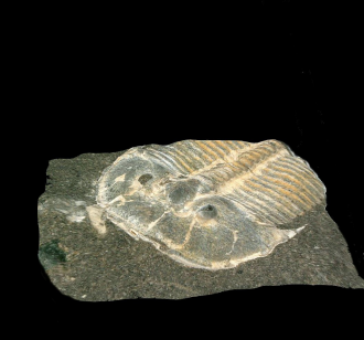
The internal structure of a 429-million-year-old fossilized trilobite eye is almost identical to that of modern bees, according to a study published in Scientific Reports. The findings suggest that the principles of vision in many insects and crustaceans today are at least half a billion years old.
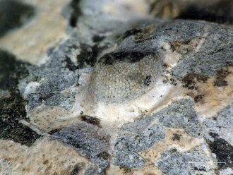
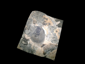
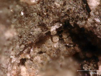
Brigitte Schoenemann and Euan Clarkson used digital microscopy to re-examine a fossilized trilobite (Aulacopleura koninckii) that was discovered in 1846 near Loděnice, Czech Republic. The fossil is 1–2 millimetres high,with two protruding semi-oval eyes on the back of its head, one of which has broken off. The authors report a number of internal structures that are similar to those of the compound eyes of many modern insects and crustaceans, including their visual units known as ommatidia (measuring 35 micrometres in diameter) that contain light-detecting cells grouped around a transparent tube called a rhabdom. The authors propose that a dark ring surrounding each individual visual unit is made from pigment cells that acted as barriers between them. Each visual unit is topped with a thick lens and the remains of what the authors suggest is a flat crystalline cone that light passed through before being focused onto the rhabdom.
The small size of its visual units indicates that A. koninckii lived in bright, shallow waters and was probably active during the day, as smaller diameter lenses are efficient at capturing light under bright conditions. The presence of pigment cell barriers between visual units suggests that the trilobite had mosaic vision with each visual unit contributing a small portion of the overall image, similar to the compound eyes of many modern insects and crustaceans.
The findings suggest that the structure and function of many compound eyes has remained mostly unchanged since the Palaeozoic era (542–251 million years ago) and provide insights into the life of an ancient trilobite.