
 Australia; WA
News release
From:
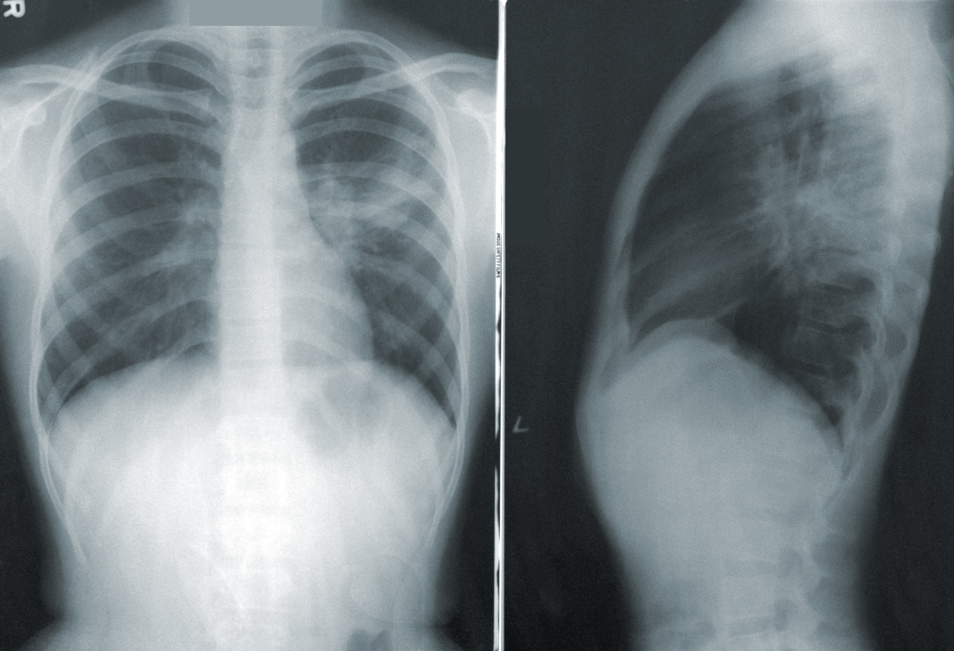
New research using machine learning on images of everyday items is improving the accuracy and speed of detecting respiratory diseases, reducing the need for specialist medical expertise.
Edith Cowan University (ECU) researchers trained algorithms on a database of more than 1 million commonplace images and transferred this knowledge to identify characteristics of medical conditions which can be diagnosed with an x-ray.
Results of this technique, known as transfer learning, achieved a 99.24 per cent success rate when detecting COVID-19 in chest x-rays.
The study tackles one of the biggest challenges in image recognition machine learning: algorithms needing huge quantities of data, in this case images, to be able to recognise certain attributes accurately.
ECU School of Science researcher Dr Shams Islam said this was incredibly useful for identifying and diagnosing emerging or uncommon medical conditions.
“Our technique has the capacity to not only detect COVID-19 in chest x-rays, but also other chest diseases such as pneumonia. We have tested it on 10 different chest diseases, achieving highly accurate results,” he said.
“Normally, it is difficult for AI-based methods to perform detection of chest diseases accurately because the AI models need a very large amount of training data to understand the characteristic signatures of the diseases.”
“The data needs to be carefully annotated by medical experts, this is not only a cumbersome process, it also entails a significant cost.”
“Our method bypasses this requirement and learns accurate models with a very limited amount of annotated data.”
“While this technique is unlikely to replace the rapid COVID-19 tests we use now, there are important implications for the use of image recognition in other medical diagnoses,” he said.
Taking a shortcut on training
Lead author and ECU PhD candidate Fouzia Atlaf said the key to significantly decreasing the time needed to adapt the approach to other medical issues was pretraining the algorithm with the large ImageNet database.
“ImageNet is a database of more than 1 million images which has been classified by humans – just like chest x-rays by medical professionals would need to be,” she said.
“The difference is the images in the database are of regular household items which can be classified by people without medical expertise.”
Dr Islam and Ms Altaf hope the technique can be further refined in future research to increase accuracy and further reduce training time.
The research paper, A novel augmented deep transfer learning for classification of COVID-19 and other thoracic diseases from X-rays was published in Neural Computing and Applications and can be accessed at the Journal’s webpage.
Attachments
Note: Not all attachments are visible to the general public. Research URLs will go live after the embargo ends.


