
 Australia; QLD
News release
From:
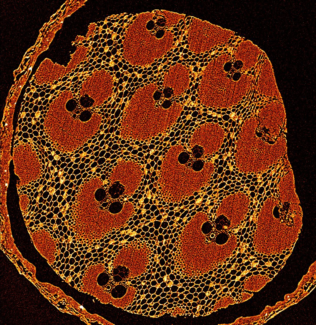
Compelling images of everyday items and intricate biological structures have revealed the capabilities of The University of Queensland’s industrial micro-CT scanner.
Associate Professor Gary Cowin, National Imaging Facility Fellow at UQ’s Australian Institute for Bioengineering and Nanotechnology (AIBN) said the 7-tonne scanner offers huge scope for use in research across both academia and industry.
“The level of detail that can be achieved is incredible, from visualising the internal structure of a toothpick to mapping the tiny brains of insects,” Dr Cowin said.
“These images demonstrate the scientific and commercial potential of this technology.”
The Yxlon FF35 micro-CT scanner can handle tiny samples, down to 1mm, or as large as 400mm.
Dr Cowin said the technology supports disciplines as diverse as engineering, biology, archaeology and materials science.
“Agricultural researchers have used the micro-CT scanner to study root growth under different soil conditions,” he said.
“Materials scientists and engineers have used it to assess porosity and cracks in carbon fibre for advanced manufacturing and verify the accuracy of 3D-printed components.
“We’ve scanned everything from mining ore samples to praying mantis brains.”
Dr Cowin said among the more striking outputs are colour-enhanced scans of native flowers and the timber of a matchstick, revealing hidden cellular structures in vivid detail.
“Being able to zoom in at the micron level allows you to see patterns and textures that are both scientifically valuable and visually interesting,” he said.
The scanner is available for use by researchers at UQ and elsewhere, as well as being accessible to commercial partners through the national facility.
“We can provide scanning services or train users to operate the system themselves, whether they’re developing new materials, investigating biological systems or need high-resolution imaging for quality control,” Dr Cowin said.
“The idea is for the technology to be as accessible as possible.”
The capability is based at UQ’s AIBN and provides a range of research capabilities including human imaging, pre-clinical MRI and molecular imaging, high resolution MRI microscopy, industrial micro-CT imaging, NMR spectroscopy, EPR Spectroscopy, radio chemistry and a cyclotron for radioisotope production.
The micro-CT scanner was funded through contributions from the National Imaging Facility (NIF) which is part of the Australian Government’s National Collaborative Research Infrastructure Strategy (NCRIS), the Queensland State Government and The University of Queensland.
Researchers and companies interested in using the micro-CT scanner are invited to contact the National Imaging Facility-Qld at AIBN.

