
 Australia; International; VIC
News release
From:
BRITISH JOURNAL OF OPHTHALMOLOGY
Externally peer reviewed? Yes
Evidence type: Observational
Subjects: People
Difference between retina’s biological age and person’s real age linked to heightened death risk
‘Retinal age gap’ potential use as screening tool, suggest researchers
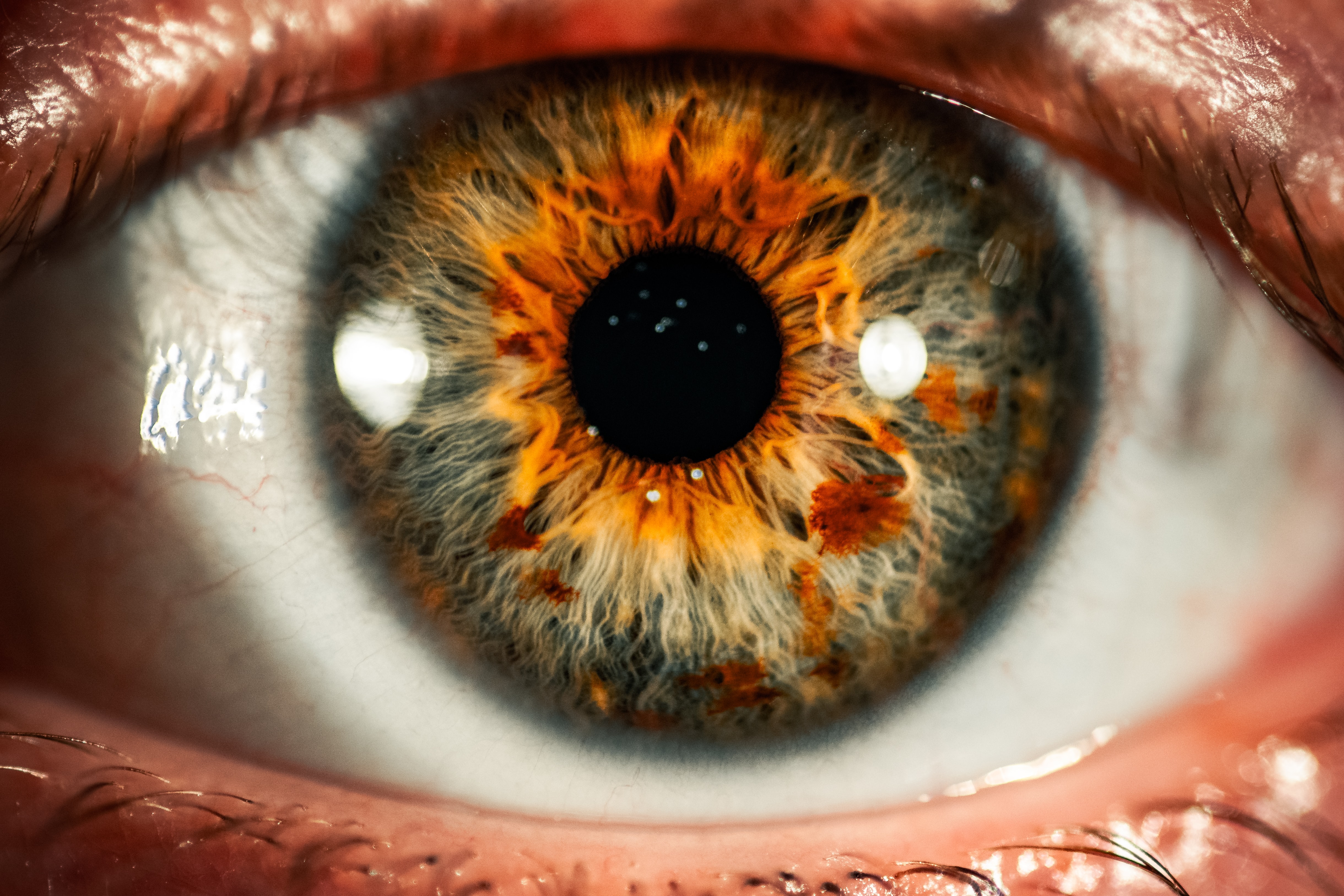
The difference between the biological age of the retina, the light sensitive layers of nerve tissue at the back of the eye, and a person’s real (chronological) age, is linked to their risk of death, finds research published online in the British Journal of Ophthalmology.
This ‘retinal age gap’ could be used as a screening tool, suggest the researchers.
A growing body of evidence suggests that the network of small vessels (microvasculature) in the retina might be a reliable indicator of the overall health of the body’s circulatory system and the brain.
While the risks of illness and death increase with age, it’s clear that these risks vary considerably among people of the same age, implying that ‘biological ageing’ is unique to the individual and may be a better indicator of current and future health, say the researchers.
Several tissue, cell, chemical, and imaging-based indicators have been developed to pick up biological ageing that is out of step with chronological ageing. But these techniques are fraught with ethical/privacy issues as well as often being invasive, expensive, and time consuming, say the researchers.
They therefore turned to deep learning to see if it might accurately predict a person’s retinal age from images of the fundus, the internal back surface of the eye, and to see whether any difference between this and a person’s real age, referred to as the ‘retinal age gap’, might be linked to a heightened risk of death.
Deep learning is a type of machine learning and artificial intelligence (AI) that imitates the way people acquire certain types of knowledge. But unlike classic machine learning algorithms that are linear, deep learning algorithms are stacked in a hierarchy of increasing complexity.
The researchers drew on 80,169 fundus images taken from 46,969 adults aged 40 to 69, all of whom were part of the UK Biobank, a large, population-based study of more than half a million middle aged and older UK residents.
Some 19,200 fundus images from the right eyes of 11,052 participants in relatively good health at the initial Biobank health check were used to validate the accuracy of the deep learning model for retinal age prediction.
This showed a strong association between predicted retinal age and real age, with an overall accuracy to within 3.5 years.
The retinal age gap was then assessed in the remaining 35,917 participants during an average monitoring period of 11 years.
During this time, 1871(5%) participants died: 321(17%) of cardiovascular disease; 1018 (54.5%) of cancer; and 532 (28.5%) of other causes including dementia.
The proportions of ‘fast agers’--those whose retinas looked older than their real age– with retinal age gaps of more than 3, 5, and 10 years were, respectively, 51%, 28%, and 4.5%.
Large retinal age gaps in years were significantly associated with 49%-67% higher risks of death, other than from cardiovascular disease or cancer.
And each 1 year increase in the retinal age gap was associated with a 2% increase in the risk of death from any cause and a 3% increase in the risk of death from a specific cause other than cardiovascular disease and cancer, after accounting for potentially influential factors, such as high blood pressure, weight (BMI), lifestyle, and ethnicity.
The same process applied to the left eyes produced similar results.
This is an observational study, and as such, can’t establish cause. The researchers also acknowledge that the retinal images were captured at one moment in time, and that the participants may not be representative of the UK population as a whole.
Nevertheless, they write: “Our novel findings have determined that the retinal age gap is an independent predictor of increased mortality risk, especially of non-[cardiovascular disease]/ non-cancer mortality. These findings suggest that retinal age may be a clinically significant biomarker of ageing.”
They add: “The retina offers a unique, accessible ‘window’ to evaluate underlying pathological processes of systemic vascular and neurological diseases that are associated with increased risks of mortality.
“This hypothesis is supported by previous studies, which have suggested that retinal imaging contains information about cardiovascular risk factors, chronic kidney diseases, and systemic biomarkers.”
The new findings, combined with previous research, add weight to “the hypothesis that the retina plays an important role in the ageing process and is sensitive to the cumulative damages of ageing which increase the mortality risk,” they explain.
Attachments
Note: Not all attachments are visible to the general public. Research URLs will go live after the embargo ends.


