
 International
News release
From:
Investigating pregnancy-related brain changes
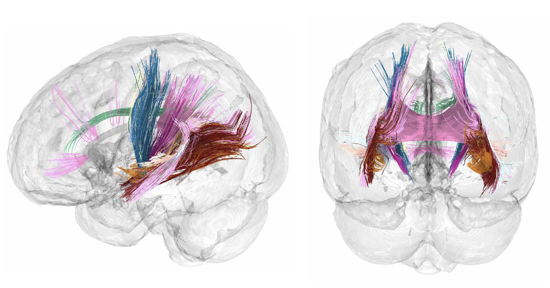
Regions of the human brain may shrink in size during pregnancy, but have better connectivity, with only a few regions of the brain remaining untouched by the transition to motherhood, according to research published in Nature Neuroscience. The findings, based on brain scans from one mother, may represent one of the first comprehensive maps of neuroanatomical changes before, during and after human pregnancy.
Nearly 85% of women become pregnant at least once during their lifetime, and 140 million women each year become pregnant. Pregnancy is known to cause physiological changes to the body, but the corresponding neural changes are not well understood.
Laura Pritschet and colleagues analysed the pregnancy-related brain changes of a healthy 38-year-old woman. They conducted 26 MRI scans and blood evaluations from 3 weeks pre-conception (4 scans) through the 3 trimesters of pregnancy, (15 scans) to 2 years postpartum (7 scans), when the testing period ended. These scans were compared to brain changes observed in 8 control individuals. The authors found widespread decreases in cortical volume and thickness by the ninth week of pregnancy, particularly in regions such as the default mode network, which is associated with social cognition. They also observed increases in white matter microstructure, ventricular volume and cerebrospinal fluid. These changes were associated with rising estradiol and progesterone hormone levels, with some persisting after birth. Some of these changes persisted at two years postpartum, including reduced cortical volume and thickness, whilst others returned to similar levels as preconception by around two months after the birth.
Although further research is needed to investigate the longer-term impacts of pregnancy on the brain, and the consistency of these brain changes across a broader population, the findings improve our understanding of the neural changes associated with pregnancy. There are also potential implications for perinatal mental health (such as neurological effects tied to pre-eclampsia or oedema), parenting behaviours and brain aging, the authors suggest.
Please note that an online press briefing for the paper below will take place UNDER STRICT EMBARGO on Thursday 12th September at 3pm London time (BST) / 10am US Eastern Time.
Authors Elizabeth Chrastil and Emily Jacobs will discuss the research. This will be followed by a Q&A session.
To attend this briefing you will need to pre-register by following the link here. Once you are registered, you will receive an email containing the details for the briefing. You will also be provided with the option to save the details of the briefing to your calendar.
Multimedia

Attachments
Note: Not all attachments are visible to the general public. Research URLs will go live after the embargo ends.


