
 International
News release
From:
A massively multi-scale approach to characterising tissue architecture by synchrotron micro-CT applied to the human placenta
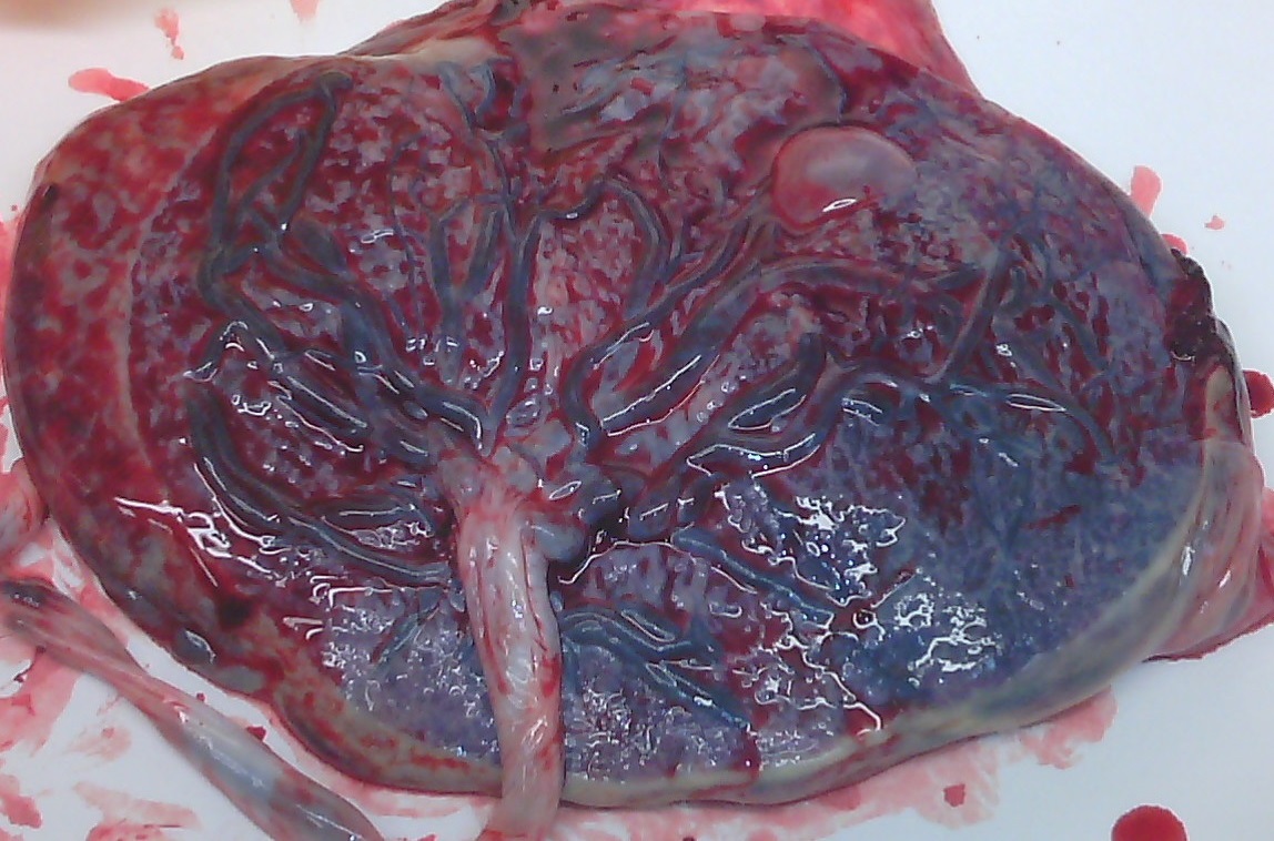
The human placenta is a critical life-support system for a rapidly developing fetus that determines their life-long health. The placenta is also one of the most complex vascular organs, where the total length of blood vessels reaches over 50 kilometres, and we still lack complete understanding how the structure of the organ determines its function. This study uses ultra-bright synchrotron X-rays, artificial intelligence and mathematical modelling to visualise and analyse human placental tissue at the unprecedented level of details. The developed tools will guide future investigations and therapies of complicated pregnancies for mothers and their babies.
Placenta mapped – The structure of the placenta, one of the most complex vascular organs, has been visualised and analysed in unprecedented detail. This study, led by the UK’s Diamond Light Source, used ultra-bright synchrotron X-rays, artificial intelligence and mathematical modelling to study the critical life-support system of the foetus. The tools developed “will guide future investigations and therapies of complicated pregnancies for mothers and their babies” the researchers said.
Attachments
Note: Not all attachments are visible to the general public. Research URLs will go live after the embargo ends.


