Cuttlefish camouflage is more complex than we thought

Publicly released:
International
Detail of the head of a cuttlefish (Sepia officinalis). CREDIT: Stephan Junek, Max Planck Institute for Brain Research.

Cuttlefish camouflage is more complex than we thought, according to international researchers who say the system is way more flexible and adaptable than previous research suggested. The team looked at the cuttlefish camouflage behaviour over natural and artificial backgrounds, gathering over 200,000 images that they used to map the colour-change process at a single-cell resolution. They found the same background could yield a multitude of different outcomes, as the cells continuously changed and corrected, which indicates the process is highly adaptable rather than a set process that happens the same way every time. The team say the only exception to this rule is ‘blanching’, a rapid and direct process where cuttlefish turn pale in response to a threat.

Media release

From: Springer Nature

Neuroscience: The complexities of cuttlefish camouflage 

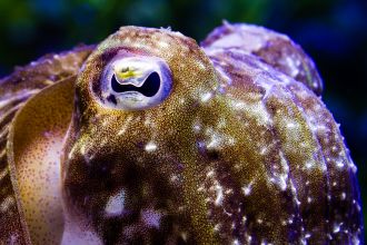
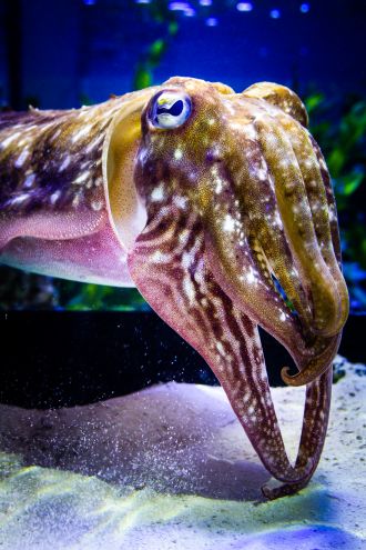
The mechanisms with which cuttlefish are able to camouflage themselves, through the control of millions of pigmented skin cells, are more complex than previously thought. The observations, published in Nature, suggest that the system is highly flexible and adaptable, providing new insights into this complex physiological process.

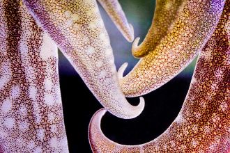
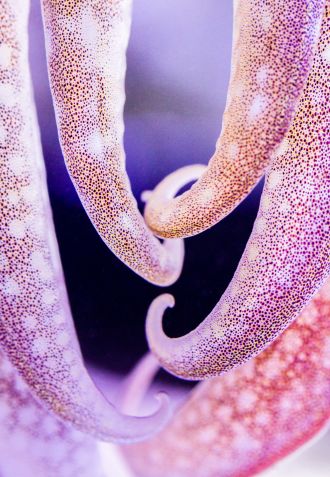
Many cephalopod species are able to camouflage themselves by matching their appearance to their surroundings. This involves a motor system that controls the expansion of several million pigment cells within the skin, known as chromatophores. The generation of skin patterns is dependent on the instinctive coordination of thousands of motor neurons that interpret complex visual scenes, a mechanism of which we have little understanding.

Gilles Laurent and colleagues studied camouflage behaviour in the common cuttlefish (Sepia officinalis) over natural and artificial backgrounds, gathering over 200,000 images that were used to map the colour-change process at a single-cell resolution. Data from these maps indicated that each pattern was highly detailed and that the same background could yield a multitude of different outcomes. These pathways to camouflage were found to involve a form of continuous feedback and the final camouflage was the product of ‘successive error-correction steps’, which indicates that the process is highly adaptable and does not follow a set path each time. The exception to this rule was during ‘blanching’, a defence mechanism in which cephalopods turn pale in response to threatening stimuli. This process was observed to be rapid and direct, and memory retained of the initial camouflage was expressed again once the threat was withdrawn. 

These results provide valuable insight into the way that these survival mechanisms interact with one another, and how the complex process of colour-matching is achieved at a cellular level.

Multimedia

Image 1: Detail of the head of a cuttlefish (Sepia officinalis).
Image 2: Head of a cuttlefish (Sepia officinalis).
Image 3: Details of the arms of a cuttlefish (Sepia officinalis).
Image 4: Details of the arms of a cuttlefish (Sepia officinalis).
Video 1
Video 2

Attachments

Note: Not all attachments are visible to the general public. Research URLs will go live after the embargo ends.

Research Springer Nature, Web page The URL will go live after the embargo lifts.
Journal/
conference:
Nature
Research:Paper
Organisation/s: Max Planck Institute for Brain Research, Germany
Funder: Open access funding provided by Max Planck Society. This research was funded by the Max Planck Society (to G.L.) and the Loewe-Schwerpunkt 2022 Center for Multiscale Modeling in the Life Sciences (to G.L.). S.R. was funded by the Okinawa Institute of Science and Technology and Kakenhi grants 60869155 and 20K15939; and D.A.E. by HFSP and EMBO long-term fellowships.
Media Contact/s
Contact details are only visible to registered journalists.