Australia; NSW
Media release
From:
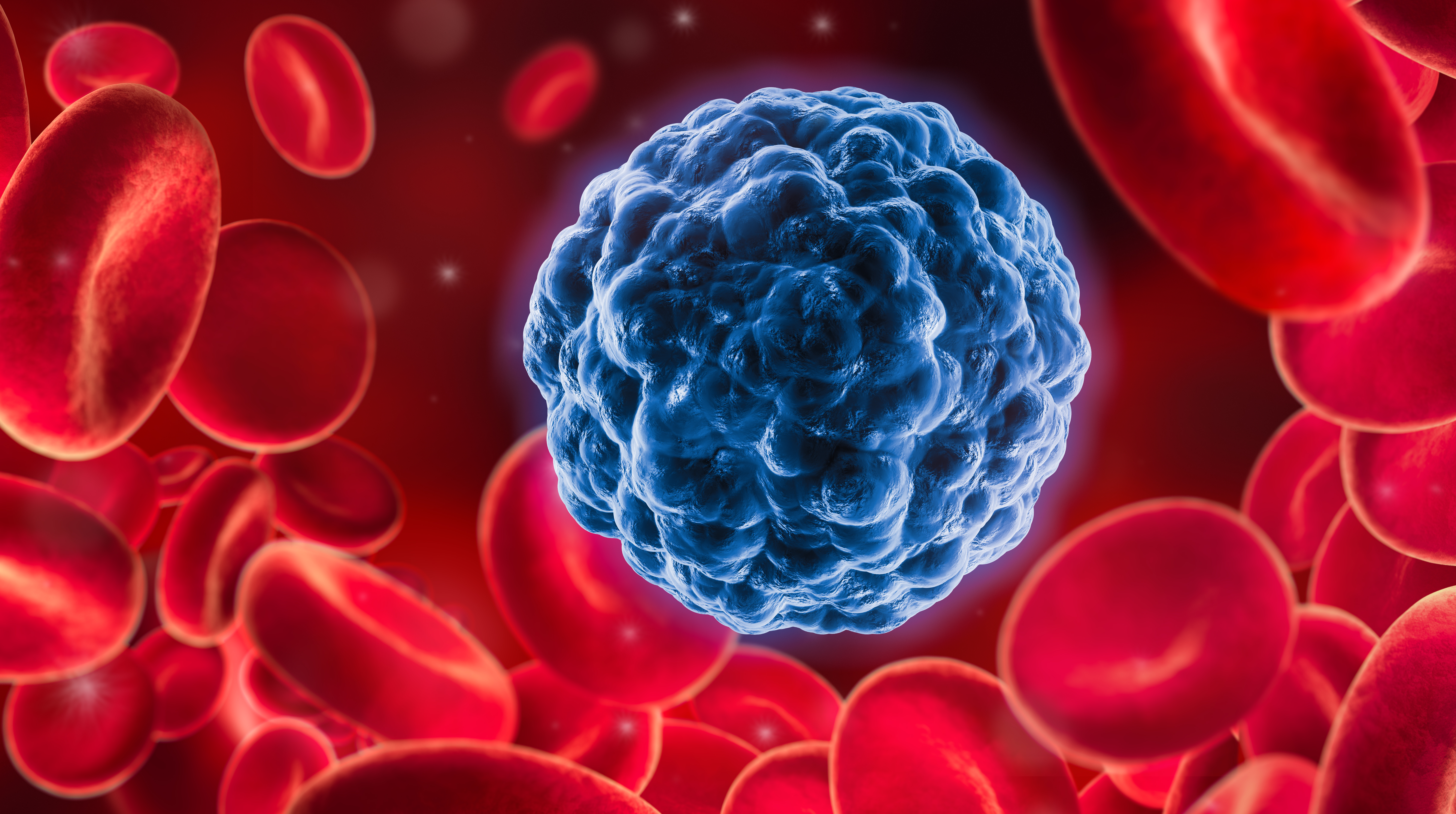
Nine of the 10 most common cancer deaths in Australia are caused by solid tumours, but in most cases it’s the cancer’s spread to other parts of the body – known as metastasis – that proves fatal.
Now, UNSW Sydney researchers have uncovered a potential trigger for metastasis: the squeezing of cancer cells by the tiniest of veins that transforms them into a different type of cell now able to form new tumours.
In a study published recently in Nature Communications, the scientists described how they constructed a biomedical device that simulated blood flow through our narrowest blood veins. They showed that when human melanoma cancer cells are forced through channels narrower than 10 micrometres – about a fifth the width of a human hair – they begin to behave more like stem cells, gaining traits that could help them survive, spread, and form new tumours.
The finding supports a theory long held by medical researchers that the mechanical pressure of narrow blood vessels might make cancer cells more aggressive. While the recent results were observed in bioengineered devices in the lab and in mice, they offer a new perspective that could inform further research and future strategies to prevent cancer from spreading.
“For a long time we’ve not fully understood how cancer spreads to distant organs,” says co-author Professor Kris Kilian, with UNSW School of Materials Science and Engineering and School of Chemistry.
“Most tumour cells circulating in blood are ill equipped to survive and spread to other organs, but we still see high rates of metastasis in some patients.
“Our finding paints a new picture, where cancer cells are triggered into becoming more tumorigenic –meaning they can form new tumours – after squeezing through the narrowest of spaces, suggesting this process may precede events like bone and brain metastasis.”
Blood vessel simulation
The researchers made a device smaller than a postage stamp that mimicked the way blood travels around the human body through progressively narrower channels simulating the tiny capillaries that are present in many tissues.
Study lead author Dr Giulia Silvani created the ‘microfluidic’ device on campus at the Australian National Fabrication Facility (ANFF), a nanotechnology joint venture between UNSW, University of Sydney and UTS. She made the channels out of PDMS – a type of biocompatible, rubbery plastic – which ranged from 30 micrometres in width, down to just 5.
Next, Dr Silvani pumped a nutrient-rich solution akin to blood plasma – and containing human melanoma cells – through the device at the same flow rate of blood flowing in capillaries.
“Within 15 minutes of being squeezed through the smallest channels, we observed how the melanoma cells became physically deformed,” Dr Silvani says.
“When we analysed the cells, we detected proteins linked to cancer spread and stem cell-like behaviour — suggesting that the mechanical stress had reprogrammed them to adopt this new state.”
To test whether these squeezed cancer cells were more likely to spread in the body, the researchers injected them into mice lacking a functioning immune system – allowing the human melanoma cells to survive and grow. After 30 days, they found that mice given the squeezed cells developed significantly more tumours in the lungs, bone and brain, compared to those given ‘unsqueezed’ melanoma cells. The researchers say this implies the squeezing makes the cancer cells more aggressive and tumorigenic.
“One of the most exciting aspects of this project was the chance to study metastatic cancer cells in a way that hasn’t been possible before,” Dr Silvani says.
“Their journey through the body is so hidden, leaving little trace and making them incredibly difficult to capture in action. But we were able to recreate that journey in the lab, giving us a rare glimpse into the moment when these cells switch into their most aggressive state.
“Being able to uncover even a small part of that mystery feels like an important step towards understanding, and ultimately stopping, the spread of cancer.”
Future treatment paths
Prof. Kilian says the findings raise new ideas about potential treatment options aimed at preventing cancer spreading from tumours.
“These results open up new possibilities for prognosis and treatment, by targeting the mechanical forces that lead to metastasis,” he says.
“We could then look at the cancer cells found in a patient’s blood stream to study their susceptibility to this sort of transformation which could help us assess the risk of metastasis for that individual.
“Or we could use MRI or other imaging techniques to identify regions with high densities of micro vessels to monitor for metastasis, possibly even intervening to make it much harder for the cells to get to these small capillaries.
“The point is, it used to be thought that it was just this extremely rare type of cell that found its way from a primary tumour to a spot where they could invade. But no, in some cases it’s actually the squeezing that changes the cell into this rare type, and it could put cancer researchers in a much stronger position to devise new treatment strategies.”
Next steps
Prof. Kilian says about 90% of the work for this project was on melanoma cancer cells, and he is hopeful the same conclusions will be drawn when researchers look at the squeezing effect on free-floating cells from other cancer tumours.
“I believe we’ll find evidence that many solid tumours metastasise this way – for instance, we’re already seeing compelling evidence when we repeat these experiments with breast cancer – and I’m looking forward to testing a range of cancer cell types in the lab,” he says.
“But I’m glad we began our study with melanoma cells, since skin cancer has such high mortality rates in Australia when it spreads to other organs. It’s vitally important that we gain a better understanding of melanoma metastasis, to better treat those suffering from this deadly disease.”
For her part, Dr Silvani is keen to do more work in microfluidics to emulate how cancer spreads through the body.
“Watching this idea grow from a spark of intuition into a real discovery has been deeply inspiring,” she says.
“It’s a powerful reminder of what can be achieved when engineering and biology come together. Cells are intricate machines, and unravelling their mysteries requires precise design and innovative tools, guided by the insights biology provides.”
Multimedia