
 Australia; VIC
Media release
From:
Researchers have created the first detailed molecular map of the human bone marrow, revealing new insights that could reshape our thinking about how an incurable blood cancer grows and spreads.
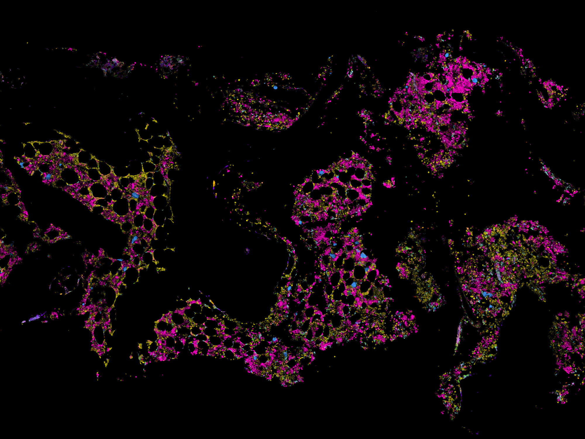
The WEHI team used state-of-the-art spatial technology to produce a molecular ‘Google map’ of the bone marrow by imaging over 5000 genes within individual cells.
The study, published in Blood, challenges established theories about the development and progression of myeloma, and paves the way for developing more effective treatments for patients.
At a glance
- First comprehensive molecular map of human bone marrow reveals surprising insights that could redefine current thinking on the development and spread of myeloma.
- The research used state-of-the-art spatial technology to create a molecular ‘Google map’, imaging over 5000 genes within individual cells.
- The findings open the door to the development of more effective myeloma treatments, tailored to individual patients.
Challenging the thinking on how myeloma behaves
Myeloma is a type of blood cancer that affects plasma cells in the bone marrow. It is often called multiple myeloma because 90% of people have multiple bone lesions when they are diagnosed.
Existing treatments can slow its progress and manage symptoms but myeloma remains incurable, with over 2500 Australians diagnosed each year.
Scientists have long believed that myeloma cells shape the bone marrow in similar ways and that universal treatments could be developed to target those common features.
But the new research reveals that each cancer cell can form their own unique microenvironment within the bone marrow.
Co-first author Dr Raymond Yip said the extraordinary level of detail in the molecular map offered fresh ways of thinking about how myeloma behaves.
“We found that each group of cancerous plasma cells creates its own distinct space, with different supporting cells and gene activity,” said Dr Yip, a postdoctoral researcher in the Hawkins Lab at WEHI.
“It’s like discovering that each tumour has its own postcode.
“Our findings challenge current thinking on myeloma and could redefine how we understand and treat the disease.
“Ultimately, this research lays the foundation for more effective treatment strategies for myeloma and potentially for other blood cancers.”
Distinct areas could explain why treatment outcomes vary
The study analysed bone marrow samples from healthy individuals, patients with early signs of disease, and those with newly diagnosed multiple myeloma.
The findings show that malignant plasma cells are not always evenly spread, but instead can cluster in spatially restricted areas, each with its own biological signature.
Clinician PhD researcher and study co-first author Jeremy Er said the research could help explain why people with myeloma respond differently to current treatments and suggests that a one-size-fits-all treatment strategy may not work.
“We hope this work is the first step in developing more tailored strategies and new ways to detect, monitor and treat myeloma,” he said.
Transforming cancer research with spatial technology
The study harnessed the latest spatial technologies at WEHI, that allow researchers to see what each cell is doing and precisely where it is located within tissue.
These powerful tools are transforming how scientists study complex diseases like cancer, by revealing how cells behave in their natural environment.
The WEHI team combined spatial transcriptomics with an optimised biobanking method for bone marrow samples, enabling them to profile 5001 genes at single-cell resolution and analyse the full cellular landscape in unprecedented detail.
The research, with collaborators from the Peter MacCallum Cancer Centre and the Royal Melbourne Hospital, was supported by the National Health and Medical Research Council (NHRMC), the Medical Research Future Fund (MRFF), Victorian Cancer Agency, Haematology Society of Australia and New Zealand (HSANZ) and Walking Up the Hill Foundation.
The team also gratefully acknowledges philanthropic support from the Roebuck Foundation and the Barrie Dalgleish Centre for Myeloma and Related Blood Cancers.
The study, “Profiling the spatial architecture of multiple myeloma in human bone marrow trephine biopsy specimens with spatial transcriptomics”, is published in Blood (DOI: 10.1182/blood.2025028896).
Multimedia

Attachments
Note: Not all attachments are visible to the general public. Research URLs will go live after the embargo ends.


