Sign In



 Australia; NSW; VIC; QLD
News release
From:
‘Invisible’ inflammation in rare bowel disease may explain heightened colon cancer risk
Researchers have mapped the cellular differences between two similar bowel diseases and identified hidden immune activity that may help explain why one carries a much higher risk of colon cancer.
New research from the Garvan Institute of Medical Research has revealed for the first time that two closely related bowel diseases, currently treated in the same way, are fundamentally different. The findings help explain why one carries a much higher risk of colon cancer – and point to the need for different approaches to treatment.
Primary sclerosing cholangitis (PSC) is a chronic liver disease affecting around 1,000 Australians that blocks the bile ducts and causes scarring of the liver. Around 70 per cent of patients also suffer from a form of inflammatory bowel disease (IBD) which resembles ulcerative colitis. Despite similarities between these conditions, people with PSC have less severe flares but face a colorectal cancer risk three times higher than those with ulcerative colitis alone. That combination – milder-looking disease with much greater cancer risk – has long puzzled researchers.
Now, findings published in Nature Communications bring us one step closer to an answer. Garvan researchers used cutting-edge methods to examine biopsies from several regions of the colon in patients with each condition, alongside healthy volunteers. The team showed that the gut of people with PSC-associated IBD had a distinct microbiome and showed a higher level of immune activity, even in the absence of inflammation, or ‘flare-ups’.
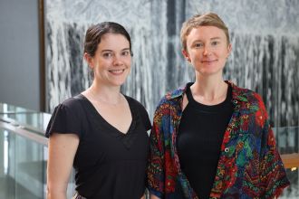
Dr Kylie James, Lab Head at the Garvan Institute and lead author of the paper says these findings open doors to more specialised treatment of PSC patients, which should ultimately lead to a reduction in colon cancer risk.
“Chronic, low-level inflammation is a known contributor to cancer risk, and our findings may explain why these patients with a milder-looking disease have a much greater chance of developing colorectal cancer. Knowing more about this disease means we can now look to develop more targeted treatments against the inflammation, even when the person isn’t having obvious symptoms of an IBD flare.”
Hidden inflammation, undetectable by current methods
The Garvan team compared biopsies from eight patients with PSC-associated IBD, 11 patients with ulcerative colitis alone, and 10 people with no gastrointestinal disease. The researchers went beyond what colonoscopy and standard biopsy examination normally show, using single-cell sequencing, spatial mapping and microbiome analysis to provide a comprehensive comparison of these conditions.
The researchers found that even in tissue that a pathologist had inspected under a microscope and classified as free from inflammation, there was a build-up of cytotoxic, or cell-killing, immune cells in the colon of patients with PSC-related bowel disease. These cells are a normal part of the immune system, but finding them so concentrated in tissue that would otherwise be considered ‘normal’ was unexpected.
“In IBD, we’ve long known that sustained remission should mean quiet immune cells, not just quiet symptoms,” says Dr James. “What we’re finding here goes further – there’s immune activity happening in tissue that a pathologist would call quiet, and it’s in the very part of the colon where these patients face their highest cancer risk. We think this constant, background inflammation could be driving the development of colorectal cancer.”
Dr Jacqueline Tearle, postdoctoral researcher at Garvan and first author of the study adds: “This is the first time this kind of immune activity has been mapped in these patients, and it tells us where to focus. If we can find ways to detect this activity in the clinic, there’s real potential to reduce the cancer risk that comes with it.”
A shared immune signal during flare-ups
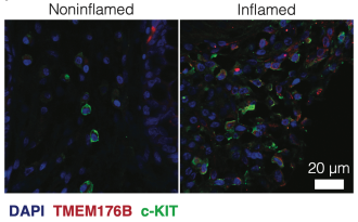
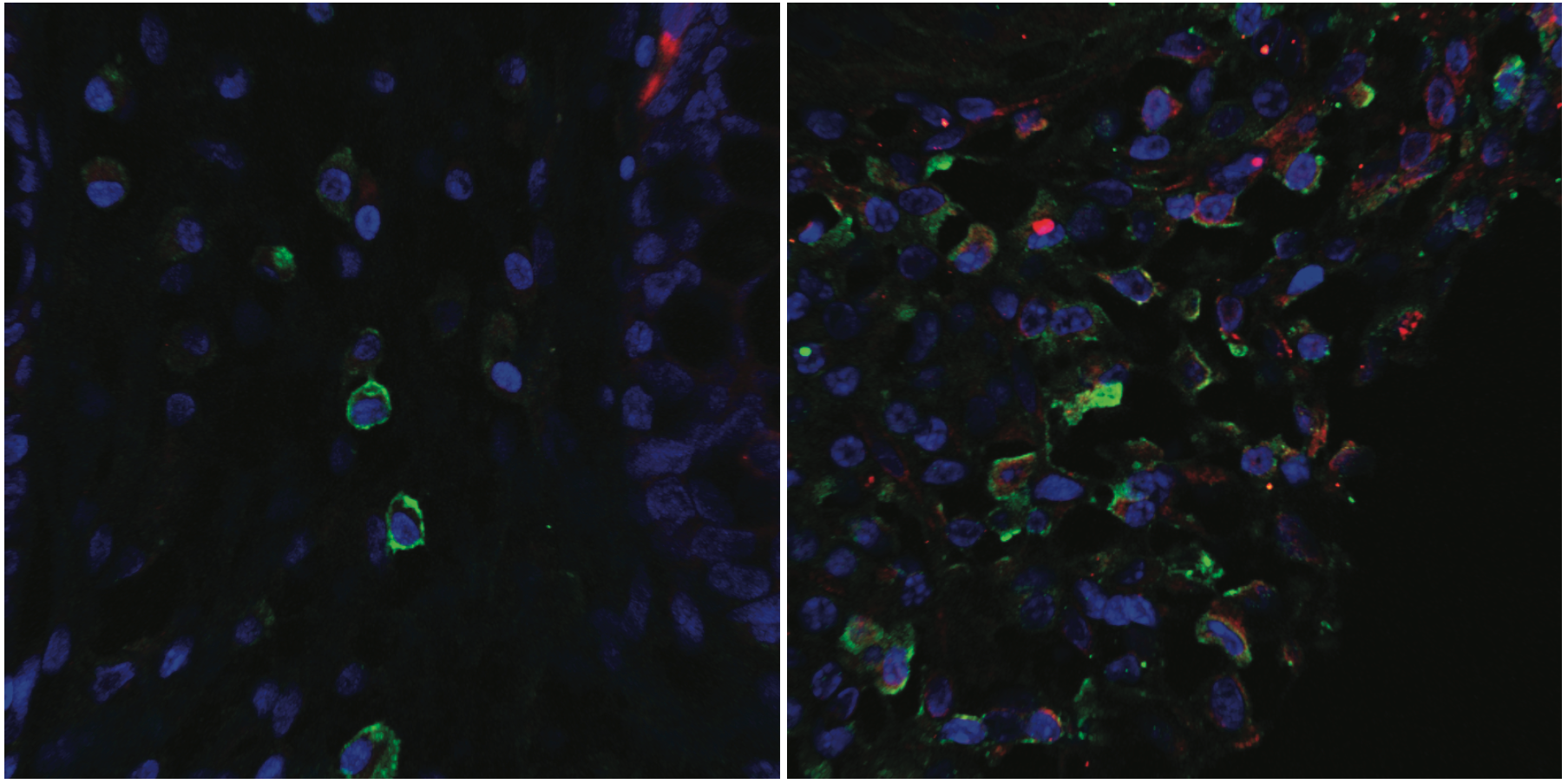
The team’s other main finding came from looking at patients during active flare-ups. In both diseases, a population of mast cells – immune cells involved in inflammation and allergic responses – built up in the gut when disease was active.
Mast cells are fragile and are typically destroyed during the standard freezing of biopsy samples, which has meant they were often overlooked in earlier studies. By processing patient tissue immediately after collection, the researchers were able to preserve them and study them in detail.
“We found quite a unique mast cell population, and its gene expression suggests it could contribute to cancer risk during periods of active inflammation or flare ups,” says Dr Tearle.
Towards treatment guided by biology
These insights could improve PSC monitoring by focusing on underlying inflammation while providing a rationale for exploring more targeted treatments.
“For patients living with PSC-related bowel disease, the risk of colorectal cancer is a real concern, and current tools don’t always capture what’s actually happening in the gut,” says Associate Professor Simon Ghaly, gastroenterologist at St Vincent’s Hospital Sydney, who led patient recruitment for the study. “Findings like these bring us closer to a more informed way of monitoring, and eventually treating, these patients.”
The team hopes that identifying these cellular features will eventually lead to treatments matched to the underlying biology of each condition, rather than to shared symptoms alone. They are now examining changes in the cells lining the colon using gut models grown from patients’ own cells, to better understand how they translate into cancer risk.
— ENDS—
This research was supported by Hillcrest Foundation, The Lumb Family, The McCusker Charitable Foundation,Australia’s National Health and Medical Research Council, The Clive and Vera Ramaciotti Foundations and the Jeremy Spinak Young Leaders Program.
Dr Kylie James is a Lab Head at Garvan Institute of Medical Research and a Conjoint Senior Lecturer at the School of Biomedical Sciences, Faculty of Medicine and Health, UNSW Sydney.
Dr Jacqueline Tearle is a Postdoctoral Research Officer at Garvan Institute of Medical Research and a Conjoint Lecturer at the School of Biomedical Sciences, Faculty of Medicine and Health, UNSW Sydney.
Associate Professor Simon Ghaly is a gastroenterologist at St Vincent’s Hospital Sydney and Conjoint Associate Professor at the School of Clinical Medicine, Faculty of Medicine and Health, UNSW Sydney.
Multimedia