
 International; NSW; VIC
News release
From:
Stem cells: Organoids created from amniotic fluid could model development in later-stage pregnancy *PRESS BRIEFING*
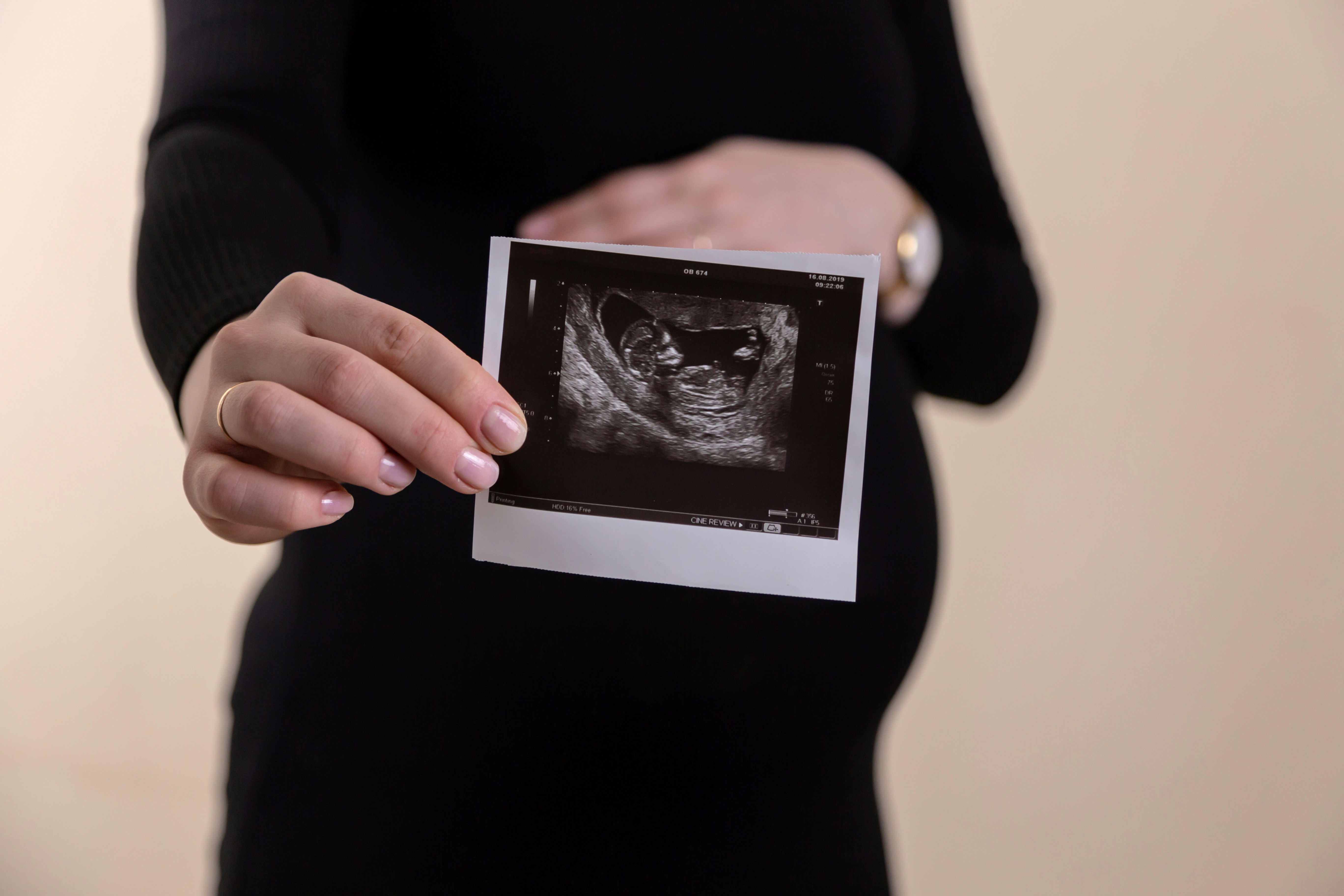
Organoids of multiple different tissue types can be generated from cells collected from amniotic fluid samples, without termination of the pregnancy, reports a study published in Nature Medicine. The organoids may provide a means of understanding development during later-stage pregnancy and could aid research into congenital anomalies.
Organoids are three-dimensional models created from human stem cells that can resemble fetal-like tissue. Current methods for deriving organoids for modeling pregnancy — mostly from post-mortem fetal tissue — have legal and ethical implications and are usually available only up to 20–22 weeks after conception, which has limited research into development in the later stages of pregnancy.
Mattia Gerli, Paolo De Coppi, and colleagues evaluated epithelial cells from human amniotic fluid collected during prenatal investigations in 12 pregnancies between the 16th week and the 34th week. Using single-cell sequencing, the authors characterised the nature of the cells and identified and isolated epithelial cells of fetal gastrointestinal, renal and pulmonary origin. To explore if these cells could be used to create organoids, the authors seeded the cells in culture and observed that the cells began proliferating and self-organising into three-dimensional organoids, visible within two weeks. They found that the cells formed tissue-specific primary fetal organoids, namely, small intestine, kidney and lungs, and that they exhibited functional features of their origin tissue. The authors were able to use this technique to generate lung organoids from amniotic and tracheal fluid cells of fetuses affected by congenital diaphragmatic hernia that recapitulated some features of this condition.
Gerli, De Coppi, and colleagues suggest that their findings demonstrate an alternative method of generating fetal organoids without termination of pregnancy that addresses long-standing ethical concerns and could be used to study later gestational stages. They indicate that it may also offer the opportunity for autologous derivation of primary fetal organoids during ongoing pregnancies, which could enable the development of advanced prenatal models and personalized therapies and help improve parental counseling. They note that further studies are needed to validate the translational impact of these findings.
***
Expert Reaction
These comments have been collated by the Science Media Centre to provide a variety of expert perspectives on this issue. Feel free to use these quotes in your stories. Views expressed are the personal opinions of the experts named. They do not represent the views of the SMC or any other organisation unless specifically stated.
Dr Evie Kendal is a bioethicist and public health scientist at Swinburne University of Technology
This research aims to close an important gap in pre-natal diagnosis and counselling for prospective parents. At present, some parents can be told their developing fetus has a disease, but not how severe it will be. This makes it very difficult for them to make informed decisions regarding potential interventions. Organoids are 3D structures developed from stem cells, the cells that contain the 'recipe' to build the different components of the body but have not been locked into a single path yet. When harvesting cells from the amniotic fluid surrounding a fetus in the womb, this research demonstrates some of these cells are committed to developing into a specific tissue type and can be used to build organoids to model the development of organs and disease.
The significance of this study includes the possibility of providing personalised counselling and targeted therapies for affected pregnancies, by helping identify which fetuses are most likely to benefit from pre-natal interventions. The method could also be used in the second and third trimester while a pregnancy continues, so fetal development could be studied at later stages of gestation than current methods allow, due to restrictions on termination of pregnancy.
From an ethical perspective, this study has the potential to improve outcomes for some future children with conditions like congenital diaphragmatic hernia, where due to malformation the abdominal organs press into the chest and compress the fetal lungs. It could enhance the autonomy of parents by providing better information to guide decision-making, as well as open areas of research in regenerative and transplant medicine that are currently closed due to ethico-legal issues with harvesting embryonic stem cells for experimentation. As organoids are not embryos, this limits potential confusion regarding their moral status.

A/Professor Kuldip Sidhu is co-founder and director, CK Cell Technologies and Conjoint with University of New South Wales Medicine.
Amniocentesis, a prenatal diagnostic test, has played a significant role in the diagnosis of foetal genetic or other chromosomal conditions since its inception, and it is generally done after 15 weeks of pregnancy. This test is available only to selected eligible pregnant women; there is a statutory ban on the use of this test for gender selection because of misuse. This test helps parents to make an informed decision about their pregnancy if the genetic report indicates a problem.
The current paper by Mattia Francesco Maria Gerl and co-workers in Nature Medicine is a state-of-the-art build on the existing technique of amniocentesis that allows scientists to do liquid biopsy of amniotic fluid for foetal genetic diagnosis.
There are many facets of this paper that exploit current cutting-edge technologies, such as single-cell sequencing, cell sorting, clonal propagation, and organoid cultures. The data as presented demonstrate a well-calibrated protocol to carry out prenatal diagnosis for foetal congenital conditions related to gastrointestinal, renal, and pulmonary stages that appear late in pregnancy.
The protocol is validated by creating an epithelial cells-derived organoid from congenital diaphragmatic hernia foetuses and exhibiting a correct diagnosis of this condition in the Petri dish. As such, it demonstrates a significant clinical progression of the existing technique that may offer respite to the perspective couples with genetic predisposition. However, the caveat is that late diagnosis of such foetal genetic diseases with this technique will add conundrum to the expected mothers and families to make difficult decisions even more difficult.
