
 Australia; QLD
Media release
From:
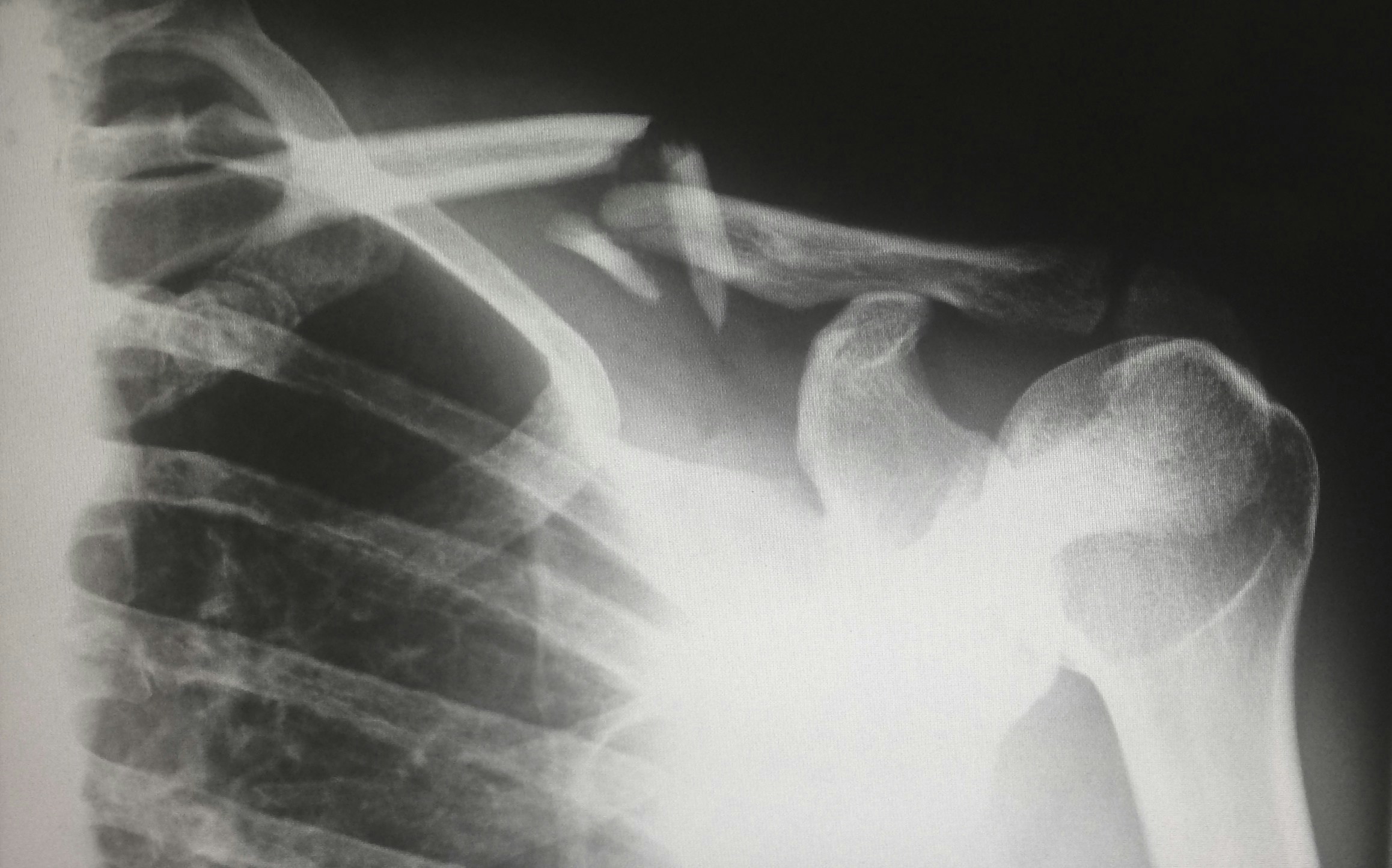
Scientists from CSIRO, Australia’s national science agency, have developed a world-first method to teach artificial intelligence (AI) how to write more accurate chest X-ray reports by giving it the same information doctors use in real life.
Using more than 46,000 real-world patient cases from a leading US hospital dataset, the team trained a powerful multimodal language model to generate detailed radiology reports.
The results showed 17 per cent better diagnostic insights and stronger alignment with expert radiologist reporting.
With hospitals worldwide struggling to keep pace with demand amid chronic radiologist shortages, this research could pave the way for faster, safer, and more reliable X-ray reporting in clinical settings.
Until now, AI tools tasked with interpreting chest X-rays relied solely on the images themselves and the doctor’s referral, without being equipped to read the vital clues hidden in patients’ medical records.
Researchers from CSIRO’s Australian e-Health Research Centre flipped that approach – combining imaging with emergency department data like vital signs, medication history and clinical notes to vastly improve diagnostic performance.
“The AI is functioning as a diagnostic detective and we’re equipping it with more evidence,” said lead author Dr Aaron Nicolson.
“When you combine what’s in the X-ray with what’s happening at the bedside, the AI gets more accurate, and much more useful.”
Dr Nicolson presented his recent findings at the international Association for Computational Linguistics conference in Vienna, Austria.
“This is a practical, scalable way to help overworked clinical teams, reduce diagnostic delays, and ultimately improve outcomes for patients,” Dr Nicolson said.
Professor Ian Scott, Research Fellow at University of Queensland Digital Health Centre and Clinical Consultant in AI at Metro South Hospital and Health Service – one of the organisations involved in testing this new technology – sees strong potential in the approach.
“For hard pressed radiologists confronting ever increasing workloads, we need this type of automated multimodal technology to reduce cognitive burden, improve workflows and allow timely and accurate reporting of chest X-rays for treating clinicians,” Professor Scott said.
Dr Nicolson and his team are currently trialling to the technology with the Princess Alexandra Hospital in Brisbane to explore how well the AI reporting compares with that of a human radiologist.
The team is also looking for other sites on which to trial the technology.
The team’s code and dataset are freely available to researchers worldwide, enabling further innovation in AI-assisted diagnostics.
Attachments
Note: Not all attachments are visible to the general public. Research URLs will go live after the embargo ends.


