
 Australia; International; VIC
Media release
From:
HEART
Externally peer reviewed? Yes
Evidence type: Observational
Subjects: People
Vascular ‘fingerprint’ at the back of the eye can accurately predict stroke risk
Combined with age and sex, predictive power as good as that of traditional risk factors alone
Practical, easily implementable approach for primary healthcare and low-resource settings
A vascular ‘fingerprint’ on the light sensitive tissue layer at the back of the eye—the retina—can predict a person’s risk of stroke as accurately as traditional risk factors alone, but without the need for multiple invasive lab tests, finds research published online in the journal Heart.
The fingerprint, comprising 29 indicators of vascular health, is a practical and readily implementable approach that is particularly well suited for primary healthcare and low-resource settings, conclude the researchers.
Stroke affects around 100 million people around the globe and kills 6.7 million of them every year, point out the researchers. Most cases are caused by modifiable risk factors, such as high blood pressure, high cholesterol, poor diet, and smoking.
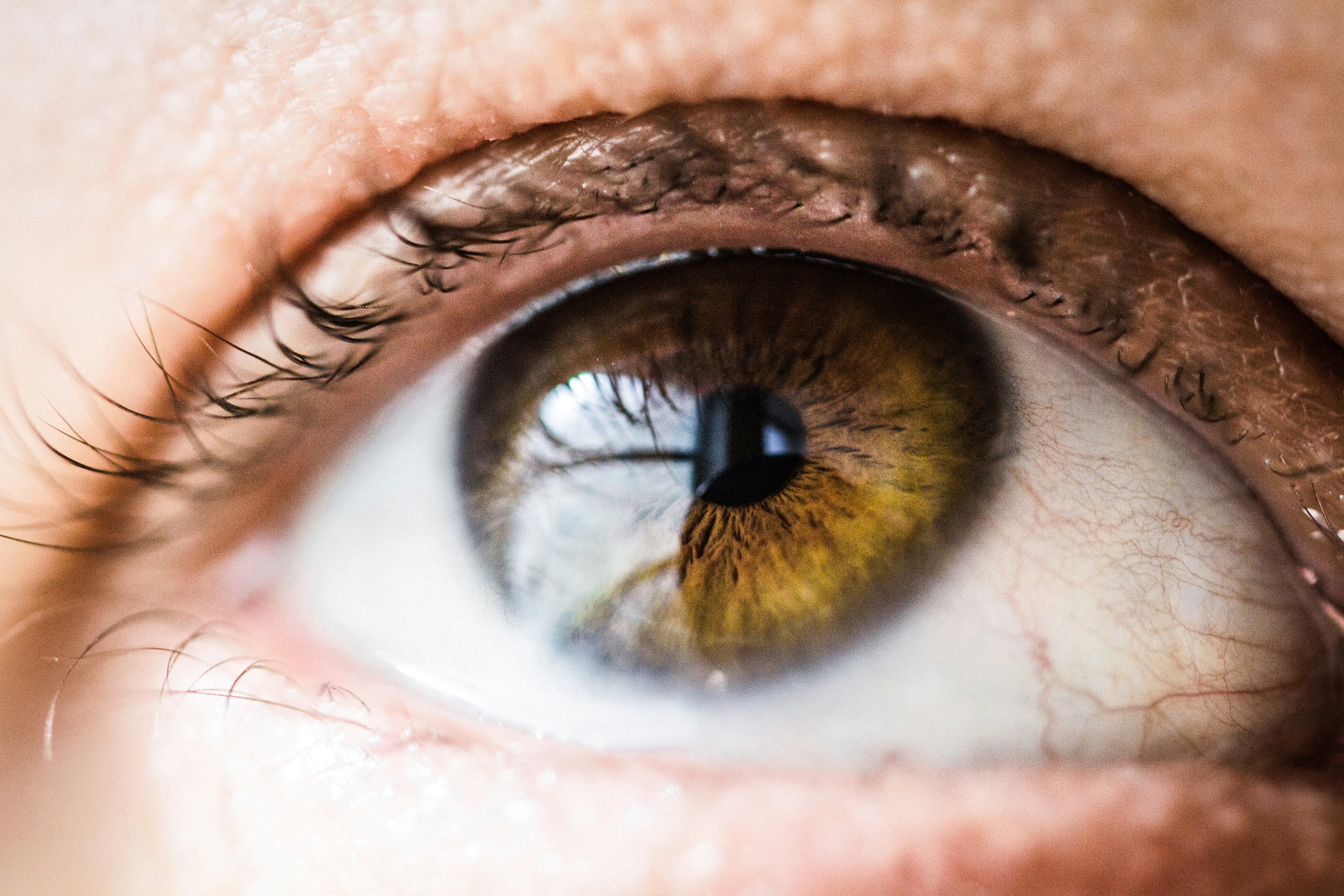
The retina’s intricate vascular network is known to share common anatomical and physiological features with the vasculature of the brain, making it an ideal candidate for assessing damage from systemic ill health, such as diabetes, explain the researchers.
Its potential for stroke risk prediction hasn’t been fully explored, due to variable study findings and inconsistent use of the specialised imaging technique for the back of the eye— fundus photography—they add.
But machine learning (AI), such as the Retina-based Microvascular Health Assessment System (RMHAS), has opened up the possibilities for the identification of biological markers that can accurately predict stroke risk without the need for invasive lab tests, say the researchers.
To explore this further, they measured 30 indicators across 5 categories of retinal vascular architecture in fundus images from 68,753 UK Biobank study participants.
The 5 categories included calibre (length, diameter, ratio) density, twistedness, branching angle and complexity of the veins and arteries.
And they accounted for potentially influential risk factors: background demographic and socioeconomic factors; lifestyle; and health parameters, including blood pressure, cholesterol, HbA1c (blood glucose indicator), and weight (BMI).
The final analysis included 45,161 participants (average age 55). During an average monitoring period of 12.5 years, 749 participants had a stroke.
These people tended to be significantly older, male, current smokers, and to have diabetes. They also weighed more, had higher blood pressure, and lower levels of ‘good’ cholesterol, all of which are known risk factors for stroke.
In all, 118 retinal vascular measurable indicators were included, of which 29 were significantly associated with first time stroke risk after adjusting for traditional risk factors. Over half (17) were density indicators; 8 fell into the complexity category; 3 were calibre indicators; and 1 came under the twistedness category.
Each change in density indicators was associated with an increased stroke risk of 10-19%, while similar changes in calibre indicators was associated with an increased risk of 10-14%.
Each decrease in the complexity and twistedness indicators was associated with an increased risk of 10.5-19.5%.
This retinal ‘vascular fingerprint’, even when combined with just age and sex, was as good as the use of traditional risk factors alone for predicting future stroke risk, the findings showed.
This is an observational study, and therefore no firm conclusions can be drawn about cause and effect. And the researchers acknowledge that the findings may not apply to diverse ethnicities as most of the UK Biobank’s participants are White. Nor were they able to assess the risk associated with different types of stroke.
Nevertheless, they conclude: “Given that age and sex are readily available, and retinal parameters can be obtained through routine fundus photography, this model presents a practical and easily implementable approach for incident stroke risk assessment, particularly for primary healthcare and low-resource settings.”
Attachments
Note: Not all attachments are visible to the general public. Research URLs will go live after the embargo ends.


