New insights into the DNA of aging hearts

Publicly released:
International
Image by Jessie Orrico on Unsplash

US researchers have identified specific mutations in human heart cells that accumulate with age. These mutations were found in the muscle cells responsible for the heartbeat and are a likely cause of many age-related heart conditions. The cumulative mutations affected the thickness of the muscle (hypertrophy), its ability to contract and relax, and in some cases caused cell loss with aging.

Media release

From: Springer Nature

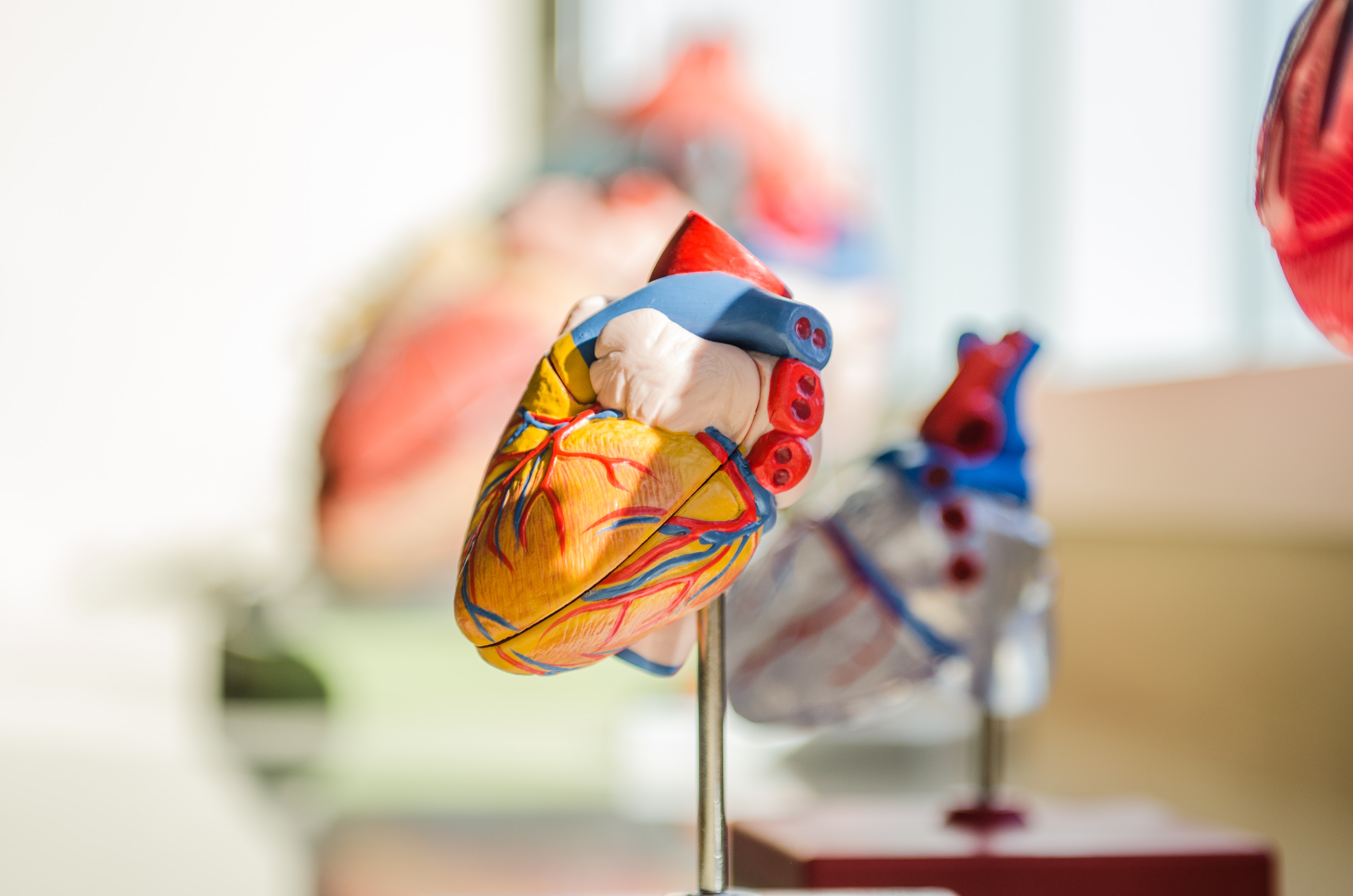
Ageing: Mutations in the ageing human heart identified

The somatic mutations that accumulate in the human heart with age are identified in a paper in Nature Aging. The findings may help us to understand how the function of the heart declines with age.

The DNA of cells that make up the human body, including the heart, accumulate errors — or somatic mutations — as we age. Although some somatic mutations appear to have little effect, others, for example, underlie the development of cancer or are likely to contribute to physiological ageing. The accumulation of somatic mutations in heart muscle cells may contribute to a decline in cellular function. However, researchers are yet to obtain data concerning these mutations.

Christopher Walsh and colleagues profile somatic single-nucleotide variants — whereby a single nucleotide in the DNA sequence is mutated — in human heart muscle cells from 12 individuals aged between 0.4 and 82 years old, using single-cell whole-genome sequencing. The authors reveal that heart muscle cells show mutational signatures that are indicative of oxidative DNA damage and failed DNA repair mechanisms, as well as an increase in base substitutions — all of which accumulate with age.

Further research will be needed to determine whether the identified processes have a causal role in heart ageing and how they may impair heart function.

Journal/
conference:
Nature Aging
Research:Paper
Organisation/s: Harvard Medical School
Funder: C.A.W. and E.A.L. are supported by the Manton Center for Orphan Disease Research and the Allen Discovery Center for Human Brain Evolution, funded by the Paul G. Allen Frontiers Program, C.A.W. is an Investigator of the Howard Hughes Medical Institute. This work is supported in part by NINDS as a supplement to R01NS032457. E.A.L is also supported by NIH (DP2AG072437; R01AG070921) and the Suh Kyungbae Foundation. E.A.M is supported by NIGMS (T32GM007753) and NLM (T15LM007092). M.B.M. is supported by NIH (K08AG065502, T32HL007627), the Brigham and Women’s Hospital Program for Interdisciplinary Neuroscience through a gift from Lawrence and Tiina Rand, the donors of the Alzheimer’s Disease Research program of the BrightFocus Foundation (A20201292F), and the Doris Duke Charitable Foundation Clinical Scientist Development Award (2021183). Authors thank the IDDRC Cellular Imaging Core, funded by NIH P50 HD105351, S10OD016453, and the Research Computing group at Harvard Medical School for assistance
Media Contact/s
Contact details are only visible to registered journalists.