
 International
News release
From:
Head trauma from sports disrupts the blood brain barrier for years after injury, a study of retired athletes shows
Traumatic head injuries from sports can disrupt the delicate blood-brain barrier and generate inflammation in athletes for more than a decade after retirement, according to a new analysis. The findings are some of the first to chart out the long-term impact of head trauma on the brain’s vascular system, and provide clues about how these disruptions may correlate with cognitive decline and other hallmarks of chronic traumatic encephalopathy (CTE).
Concussions and head injuries from sports such as American football, boxing, and rugby impact between 1.6 and 3.8 million people each year in the U.S. Rates of subconcussive injuries – those that exert forces below the concussion threshold but are still harmful – are even higher. Over time, repetitive injuries can lead to CTE, a neurodegenerative disorder that can only be diagnosed with an autopsy. Some research suggests that disruptions to the blood-brain barrier contribute to the onset of CTE, but there are no data on how sports-related head injuries affect the blood-brain barrier over the long term.
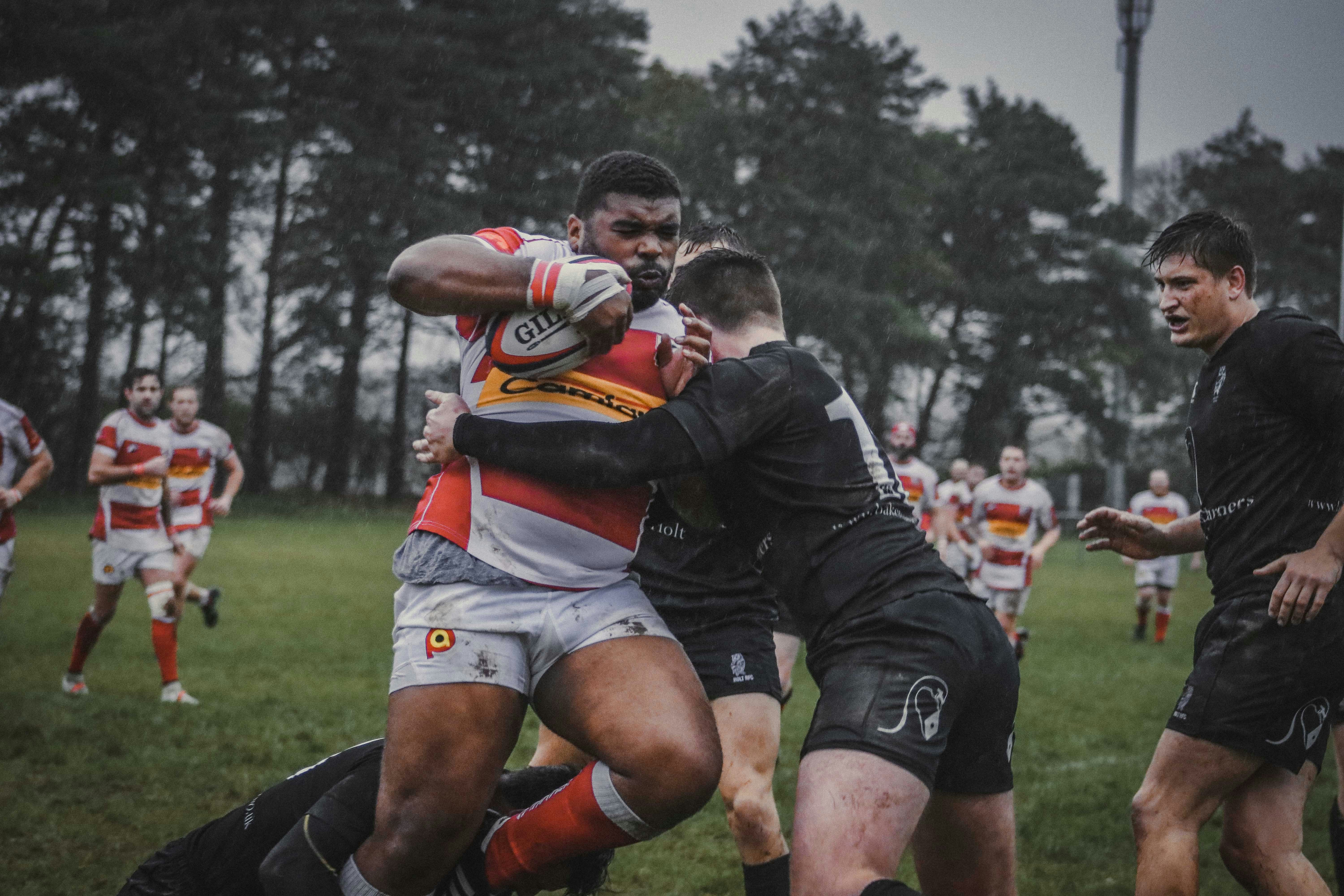
Now, Chris Greene and colleagues present one of the first analyses of the impact of sports injuries on the blood-brain barrier in athletes long after retirement. They used MRI to image the brains of 47 retired athletes, who were primarily rugby players but also included boxers, Gaelic footballers, and others. The authors also recruited controls, including non-athletes as well as athletes with backgrounds in non-contact sports such as rowing. The contact sports athletes had been retired for an average of 12.1 years. Despite this length of time, the imaging revealed clear markers of disruption to the blood-brain barrier. It also detected abnormalities in the brain’s complement system, including altered expression of the complement receptors C5AR1, ITGAM, ITGB2, and CD59.
The team also identified a subgroup of 17 athletes who not only displayed extensive leakage in the blood-brain barrier, but also showed worse cognitive performance, lower brain volumes, and elevated monocyte counts. Greene et al. note that their work is limited by its small sample size, but say it could nevertheless help identify future targets to ameliorate blood-brain barrier disruption and CTE.
Attachments
Note: Not all attachments are visible to the general public. Research URLs will go live after the embargo ends.


