Images uncover the secrets of the narwhal tusk

Publicly released:
International
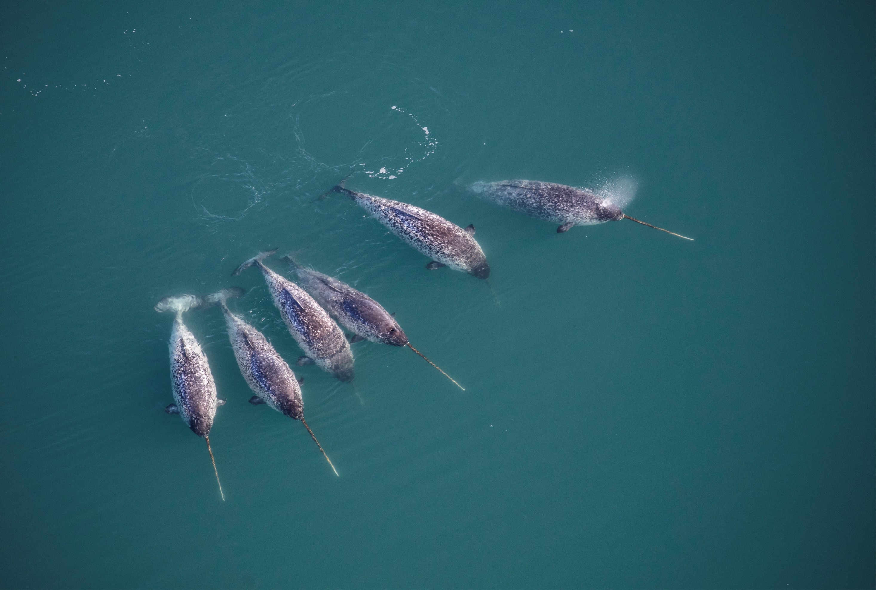
Narwhals with tusks swimming in North West Greenland. Credit: Carsten Egevang

International researchers have discovered why narwhals, the unicorns of the sea, have such straight tusks. The team combined multiple imaging techniques to figure out how narwhal tusks – which can grow to over two metres in length – grow so long and straight, as they’re the only known straight tusks found in nature. They found that, at a microscopic level, the large-scale helical (twisting) structures are produced by arrangements of mineralised collagen. However, they found the inner layer twists right, while the outer layer covering the tusk twists left, which could be what enables the narwhal tusk to grow straight while maintaining its distinctive left-handed twist.

News release

From: Springer Nature

Biology: Secrets of the narwhal tusk *IMAGES*

Hidden structures within the narwhal tusk reveal how it is able to remain so straight, according to research in Nature Communications. The findings suggest that the tusk’s distinctive left-handed spiral arises from opposing helical structures at microscopic scale.

The narwhal tusk is a protruding straight tooth that can exceed two metres in length while maintaining a characteristic left-handed spiral. The narwhal tusk is the only example of straight tusk in nature and has fascinated cultures across history. Previous studies have suggested that this shape might be linked to the organisation of the tusk’s underlying tissues, but it has remained unclear whether the macroscopic spiral structure is reflected in the tusk’s microscopic building blocks.

Henrik Birkedal and colleagues combined multiple imaging techniques to map the structure of the narwhal tusk across several scales. These observations showed that the large-scale helical structure is produced by arrangements of mineralised collagen (the building blocks of the tusk) at a microscopic level. These collagen fibrils were predominantly aligned along the length of the tusk. However, subtle but consistent changes in fibril orientation form two opposing helical arrangements: a left-handed helix in the outer cementum (the thin layer covering the tusk) and a right-handed helix in the inner dentine (an internal layer). The authors suggest that these opposing helical motifs enable the narwhal tusk to grow straight while maintaining its distinctive left-handed helix.

Further research will be needed to understand how the tusk is formed during development. However, these findings shed new light on how microscopic structural patterns can drive specific functions on a larger scale in biology.

Multimedia
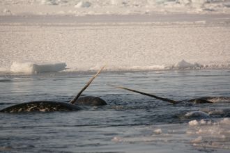
Image 1
Image 1
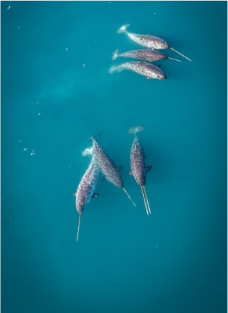
Image 2
Image 2
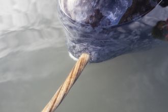
Image 3
Image 3
Image 4
Image 4

Attachments

Note: Not all attachments are visible to the general public. Research URLs will go live after the embargo ends.

Research Springer Nature, Web page The URL will go live after the embargo lifts.
Journal/
conference:
Nature Communications
Research:Paper
Organisation/s: Aarhus University, Denmark
Funder: This work is supported by NORDFORSK grant 105053, Villum Investigator grant 25867, and the Aarhus University Research Foundation NOVA grant.We thank the Danish Agency for Science, Technology, and Innovation for funding the instrument center DanScatt. Funded by the European Union. Views and opinions expressed are, however those of the author(s) only and do not necessarily reflect those of the European Union or European Research Executive Agency. Neither the European Union nor the granting authority can be held responsible for them. ARP has received funding from the European Union’s Horizon 2020 research and innovation program under the Marie Skłodowska-Curie grant agreement No 101152202, and the Area of Advance (Material Science) at Chalmers University of Technology and the Swedish Research Council (VR 2018-041449). ML and LCN received funding from the European Research Council (ERC-2020-StG 949301). CA has received funding from the European Union’s Horizon 2020 research and innovation program under the Marie Skłodowska-Curie grant agreement No 884104 and from theChalmers initiative for the advancement of neutron and X-ray techniques. Research conducted at MAX IV, a Swedish national user facility, is supported by Vetenskapsrådet (Swedish Research Council, VR) under contract 2018-07152, Vinnova (Swedish Governmental Agency for Innovation Systems) under contract 2018- 04969, and Formas under contract 2019-02496. DanMAX is funded by the NUFI grant no. 4059-00009B. The DanMAX XRF detector was funded by Carlsberg: CF18-0802. HPC2N was partially funded by the Swedish Research Council through grant agreement no. 2018-05973.
Media Contact/s
Contact details are only visible to registered journalists.