Electrical stimulation helped nine paralysed people walk again

Publicly released:
International
Adapted from Extended Figure 1 in paper. Credit: Courtine, et al. (2022)

Nine people with chronic spinal cord injuries have regained the ability to walk after being treated with epidural electrical stimulation (EES), according to a small study by international researchers. The team used EES to activate specific sets of neurons that become necessary for patients to walk after paralysis from severe or complete paralysis caused by spinal injury. All patients immediately regained or improved their ability to walk during the treatment, and showed improvements in mobility after five months of EES treatment and rehabilitation. To further explore how this worked, the team looked at similar pathways in mice and found a specific kind of excitatory neuron that plays an important role in restoration of walking after spinal cord injury, but is not necessary for walking in people without spinal cord injury.

Media release

From: Springer Nature

Neuroscience: Identifying neurons that restore walking after paralysis (N&V)

The neurons that promote recovery from paralysis are identified in a study in which nine individuals with chronic spinal cord injury regained the ability to walk after being treated with electrical stimulation. The findings, presented in a Nature paper this week, improve our understanding of how mobility can be recovered after paralysis.

Electrical stimulation of the spinal cord has been found to be effective in improving recovery of walking in people with paralysis, but the underlying mechanism of this treatment remains unclear.

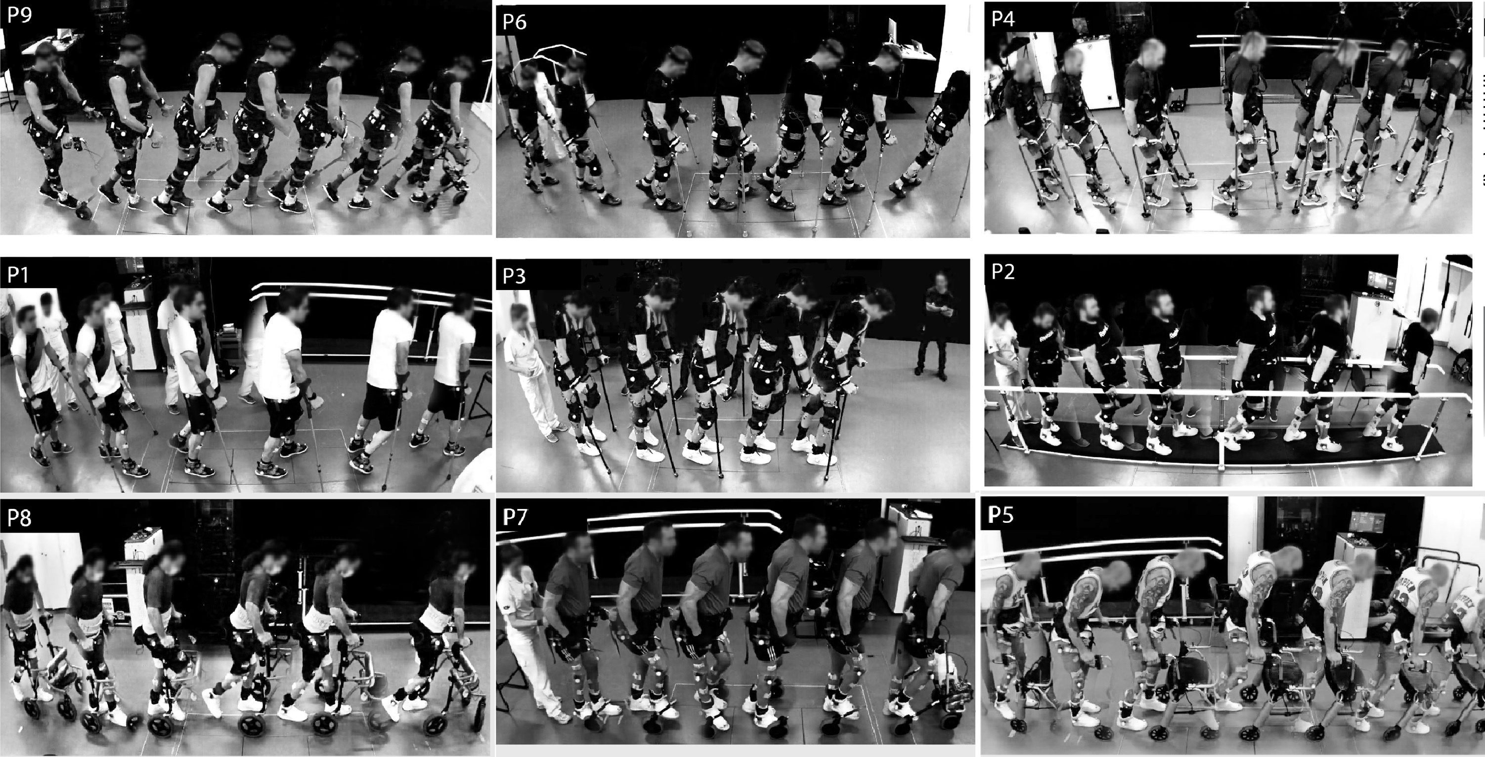
Grégoire Courtine and colleagues investigated whether electrical stimulation might recruit specific sets of neurons in the spinal cord that become necessary for patients to walk after paralysis. In this study, nine individuals with severe or complete paralysis caused by spinal cord injury were enrolled in a clinical trial and received epidural electrical stimulation (EES) treatment. All patients immediately regained or improved their ability to walk during the treatment and showed improvements in mobility after five months of EES treatment and rehabilitation. To explore the underlying mechanism of this improvement, the authors developed a mouse model that replicates the key features of EES neurorehabilitation in humans. In addition, they established a single-cell map of gene expression in various neurons of the mouse spinal cord. Combining the model and the molecular map, the authors identified a specific type of excitatory neuron that plays an important role in restoration of walking after spinal cord injury but is not necessary for walking in individuals without spinal cord injury.

The findings bring us a step closer to understanding the mechanisms of EES rehabilitation. However, the authors note that other neurons in the brain and spinal cord contribute to the recovery of walking, and therefore further studies are needed.

Attachments

Note: Not all attachments are visible to the general public. Research URLs will go live after the embargo ends.

Research Springer Nature, Web page The URL will go live after the embargo lifts.
Journal/
conference:
Nature
Research:Paper
Organisation/s: Swiss Federal Institute of Technology (EPFL), Switzerland
Funder: This work was supported by Defitech Foundation, Rolex for Enterprise, Carigest Promex, Wings for Life, Riders4Riders, ALARME, Panacée Foundation, Pictet Group Charitable Foundation, Firmenich Foundation, the Bertarelli Foundation, International Foundation for Research in Paraplegia, ONWARD medical, the Swiss National Science Foundation (National Centre of Competence in Research in Robotics, subside 51NF40_185543) and grants (310030_192558 to G.C.), InnoSuisse STIMO Bridge (41871.1 IP-LS), Eurostars E!12743 CONFIRM, Eurostars E!113969 PREP2GO, Swiss National Science Foundation (32003BE_205563), European Research Council (ERC-2015-CoG HOW2WALKAGAIN 682999; Marie Sklodowska-Curie individual fellowship 842578 to J.W.S.), H2020-MSCA-COFUND-2016 EPFL Fellows programme (no. 665667 to C.K.), Human Frontiers in Science Program long-term fellowship (LT001278/2017-L to C.K.), the Swiss National Supercomputing Center (CSCS), and the Intramural Research Program of the NIH, NINDS. M.A.S. acknowledges support from the Wings for Life Spinal Cord Research Foundation. We thank J. Ravier and M. Burri for illustrations; B. Schneider and T. Karayannis for providing viral vectors; V. Paggi for guidance on implant microfabrication; L. Batti and I. Gantar from the Advanced Lightsheet Imaging Center (ALICe) at the Wyss Center for Bio and Neuroengineering, Geneva. This work was supported in part using the resources and services of the Gene Expression Core Facility at the School of Life Sciences of EPFL.
Media Contact/s
Contact details are only visible to registered journalists.