
 Australia; WA
News release
From:
Thanks to artificial intelligence, we will soon be able to predict our risk of developing serious health conditions later in life, at the press of a button.
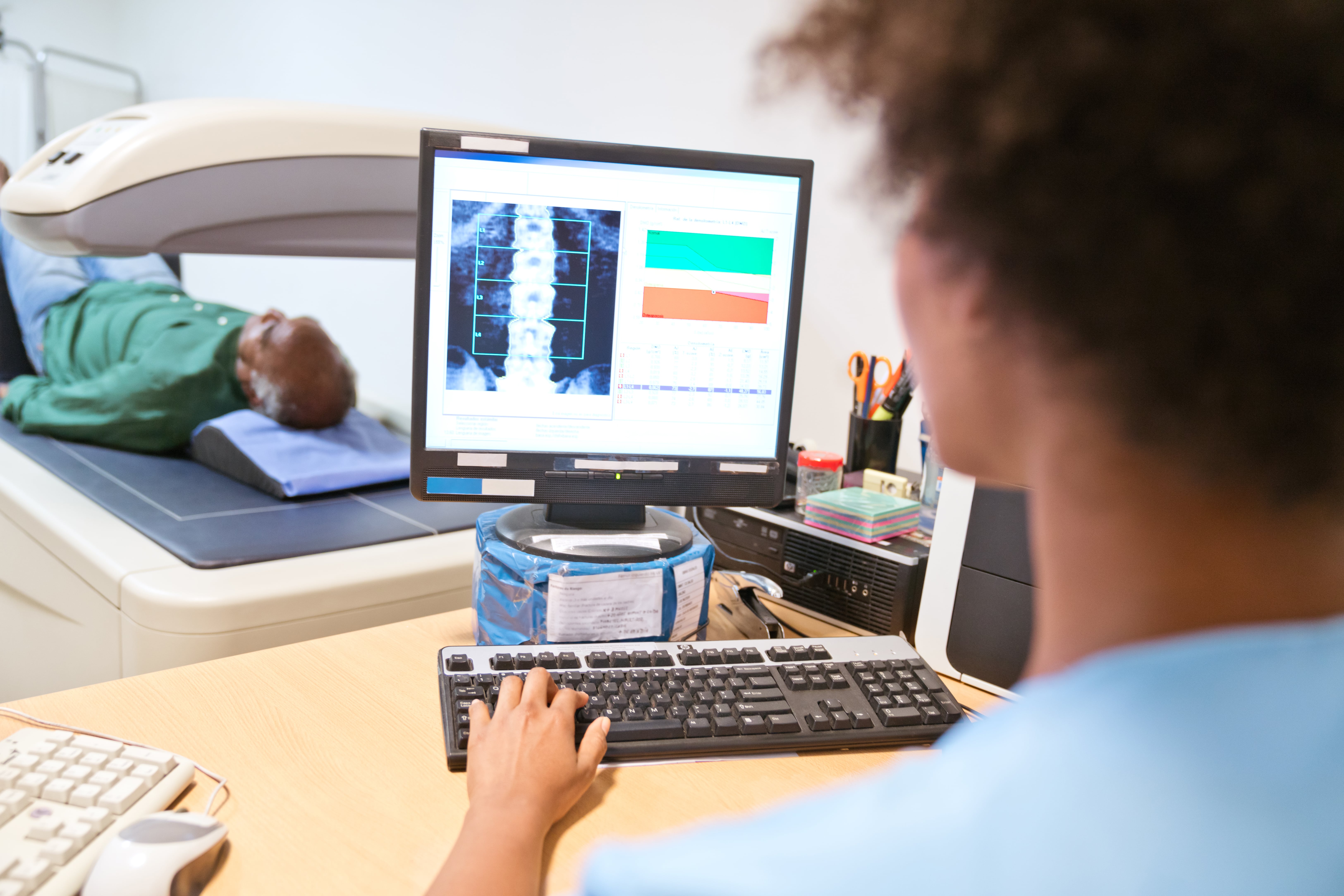
Abdominal aortic calcification, or AAC, is a calcification which can build up within the walls of the abdominal aorta and predicts your risk of developing cardiovascular disease events such as heart attacks and stroke.
It also predicts your risk of falls, fractures and late-life dementia.
Conveniently, common bone density machine scans used to detect osteoporosis, can also detect AAC.
However, highly trained expert readers are needed to analyse the images, a process which can take 5-15 minutes per image.
But researchers from Edith Cowan University’s (ECU) School of Science and School of Medical and Health Sciences have collaborated to develop software which can analyse scans much, much faster: roughly 60,000 images in a single day.
Researcher and Heart Foundation Future Leader Fellow Associate Professor Joshua Lewis said this significant boost in efficiency will be crucial for the widespread use of AAC in research and helping people avoid developing health problems later in life.
“Since these images and automated scores can be rapidly and easily acquired at the time of bone density testing, this may lead to new approaches in the future for early cardiovascular disease detection and disease monitoring during routine clinical practice,” he said.
Saving a LOT of time
The results were from an international collaboration between ECU, the University of WA, University of Minnesota, Southampton and University of Manitoba, Marcus Institute for Aging Research, and Hebrew SeniorLife Harvard Medical School. Truly a multidisciplinary global effort.
Though it’s not the first algorithm developed to assess AAC from these images, the study is the biggest of its kind, was based on the most commonly used bone density machine models, and is the first to be tested in a real-world setting using images taken as part of routine bone density testing.
It saw more than 5000 images analysed by experts and the team’s software.
After comparing the results, the expert and software arrived at the same conclusion for the extent of AAC (low, moderate or high) 80 per cent of the time – an impressive figure given it was the first version of the software.
Importantly, only 3 per cent of people deemed to have high AAC levels were incorrectly diagnosed to have low levels by the software.
“This is notable as these are the individuals with the greatest extent of disease and highest risk of fatal and nonfatal cardiovascular events and all-cause mortality,” Professor Lewis said.
“Whilst there is still to work to do to improve the software’s accuracy compared to human readings, these results are from our version 1.0 algorithm, and we already have improved the results substantially with our more recent versions.
“Automated assessment of the presence and extent of AAC with similar accuracies to imaging specialists provides the possibility of large-scale screening for cardiovascular disease and other conditions – even before someone has any symptoms.”
“This will allow people at risk to make the necessary lifestyle changes far earlier and put them in a better place to be healthier in their later years.”
The Heart Foundation provided funding for the project, thanks to Professor Lewis’ 2019 Future Leadership Fellowship providing support for research over a three-year period.
‘Machine Learning for Abdominal Aortic Calcification Assessment from Bone Density Machine-Derived Lateral Spine Images’ was published in eBioMedicine.

